. 2008 Aug 8;266(2):249-62.
doi: 10.1016/j.canlet.2008.02.062. Epub 2008 Apr 8.

Priming with EGFR tyrosine kinase inhibitor and EGF sensitizes ovarian cancer cells to respond to chemotherapeutical drugs

Affiliations

Priming with EGFR tyrosine kinase inhibitor and EGF sensitizes ovarian cancer cells to respond to chemotherapeutical drugs

Cong Cao et al. Cancer Lett. .

Abstract

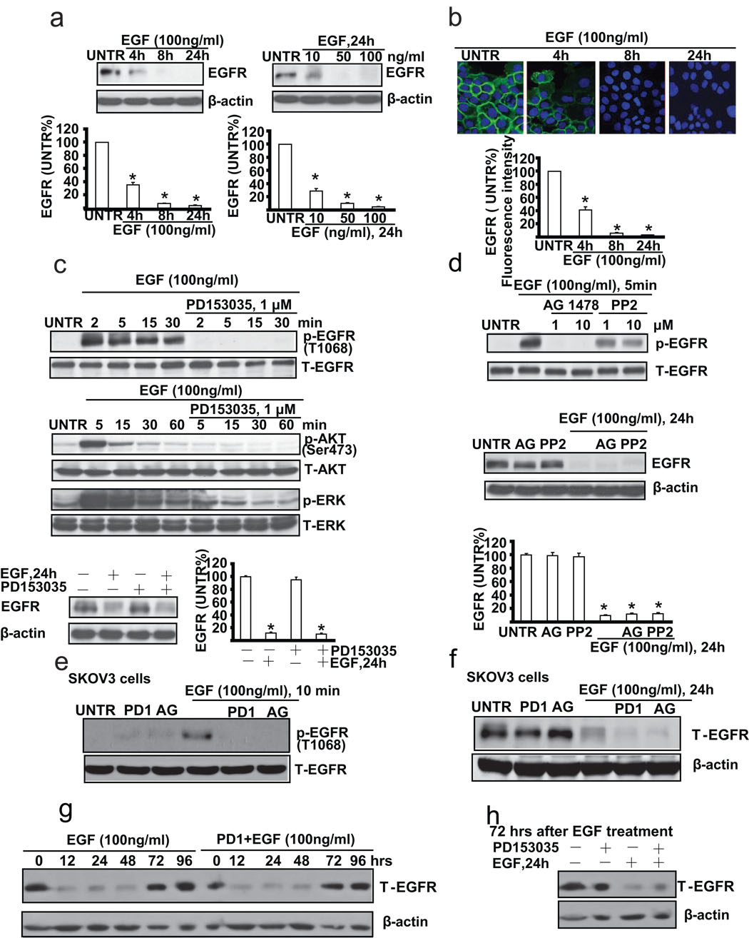
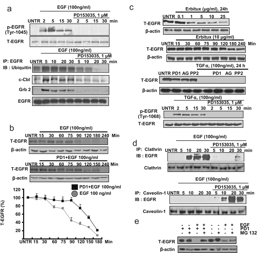
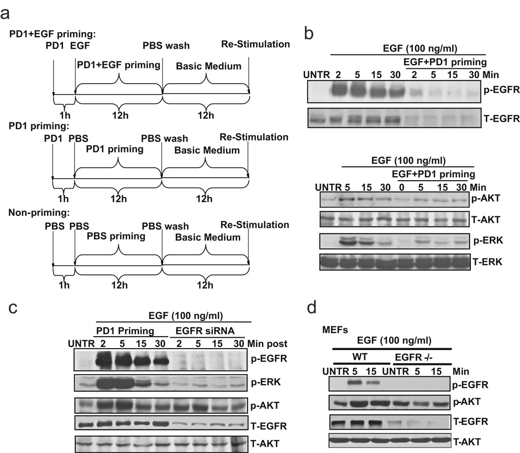
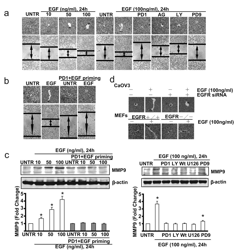
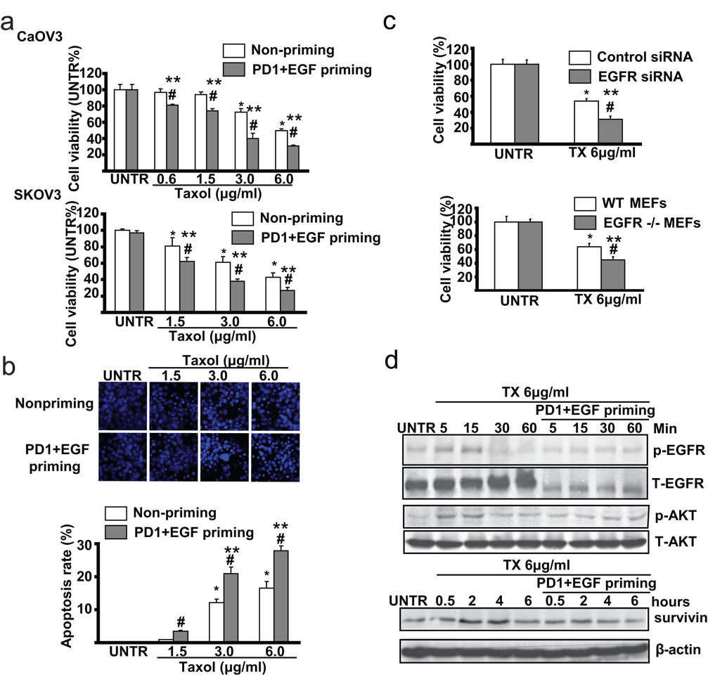
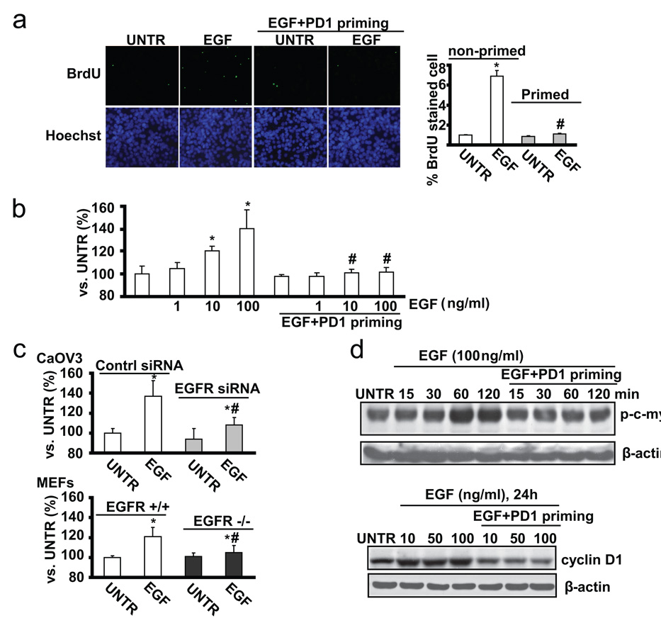
Over-expression of EGFR, as in most cases of ovarian cancer, is associated with advanced-stage disease and poor prognosis. Activation of EGFR signaling pathway is involved in increased cell proliferation, angiogenesis, metastasis and decreased apoptosis. Tyrosine kinase activity is essential for signal transduction and receptor down-regulation. However, we found in this study that tyrosine kinase activity is not necessary in ligand-induced EGFR down-regulation in ovarian cancer cell line CaOV3 cells. EGFR tyrosine kinase inhibitors, such as PD153035, AG1478, as well as non-specific tyrosine kinase inhibitor PP2 cannot reverse EGF-induced down-regulation of EGFR. These findings thus permit us to develop the following exciting but unconventional strategy to sensitize cancer cells, namely, by priming ovarian cancer cells with EGF and EGFR inhibitor PD153035, before chemotherapy. This priming procedure down-regulates EGFR without induction of mitogenic signals such as ERK and PI3K/AKT. EGF plus EGFR inhibitor-primed ovarian cancer cells display increased sensitivity to taxol-induced cell death, resistant to EGF-induced cell migration and cell proliferation as well as ERK and PI3K/AKT activation. Further studies showed that PD153035, which does not reverse ligand-induced EGFR down-regulation, blocks EGF-induced EGFR activation as well as EGFR's binding to c-cbl and Grb2. Taken together, we contend that priming with EGFR inhibitors plus EGF inhibits cell signaling pathways leading to cell proliferation and survival, while down-regulating EGFR. This priming approach sensitizes ovarian cancer cells and would ultimately result in better chemotherapeutical outcome.


Figures

Fig. 1
Fig. 1. EGFR tyrosine kinase inhibitor does not reverse EGF-induced EGFR down-regulation in cultured ovarian cancer cell line CaOV3 cells
(a) CaOV3 cells were treated with EGF (100 ng/ml), harvested at different time points (4, 8 and 24 h) or treated with various doses of EGF (10, 50, and 100 ng/ml) and harvested at 24 hours. EGFR expression was analyzed by Western blot and its expression was quantified as normalized to beta actin. (b) CaOV3 cells were treated with 100 ng/ml of EGF and EGFR expression detected by immunofluorescence method as described above was observed at different time points (4, 8 and 24 h). (c) CaOV3 cells were pretreated with EGFR inhibitor PD153035 (1 µM) for 1 hour, followed by EGF (100 ng/ml) treatment for different time as indicated. p-EGFR (Tyr1068), T-EGFR, p-AKT (Ser473), T-AKT, p-ERK, T-ERK were detected by Western blot. CaOV3 cells were also pretreated with PD153035 (1 µM) for 1 hour, followed by EGF (100 ng/ml) treatment for 24 hours. T-EGFR was detected by Western blot. EGFR expression was quantified as normalized to beta actin. (d) CaOV3 cells were pretreated with AG1478 (1 and 10 µM) or PP2 (1 and 10 µM) for 1 hour, followed by EGF (100 ng/ml) treatment for 5 minutes. p-EGFR (T-1068) and T-EGFR were detected by Western blot. CaOV3 cells were pretreated with AG1478 (AG, 1 µM) or PP2 (1 µM) for 1 hour, followed by EGF (100 ng/ml) treatment for 24 hours. T-EGFR and β-actin were detected by Western blot as normalized to beta actin. (e) SKOV3 cells were pretreated with PD153035 (PD1, 1 µM) or AG 1478 (AG, 1 µM) for 1 hour, followed by EGF (100 ng/ml) treatment for 10 minutes, p-EGFR (Tyr1068), T-EGFR were detected by Western blot. (f) SKOV3 cells were pretreated with PD153035 (PD1, 1 µM) or AG 1478 (AG, 1 µM) for 1 hour, followed by EGF (100 ng/ml) treatment for 24 hours, T-EGFR and β-actin were detected by Western blot. (g) CaOV3 cells were pretreated with or without PD 153035 (PD1, 1 µM) for 1 hour, followed by EGF (100 ng/ml) treatment for indicated time, T-EGFR and β-actin were detected by Western blot. (h) After 72 hours of original EGF treatment, CaOV3 were pretreated with or without PD153035 (PD, 1 µM) for 1 hour, followed by secondary EGF (100 ng/ml) treatment for additional 24 hours, T-EGFR and β-actin were detected by Western blot. The data represent mean ± SE of triplicate experiments. * P < 0.05 versus UNTR groups. For immnofluorescence experiment, a minimum of six random fields and 200 cells per group were selected and average intensity for each group was quantified. Magnification: (b) 1: 400.
Fig. 2
Fig. 2. Cytoplasmic tyrosine kinase is not necessary for ligand-induced EGFR down- regulation
(a) CaOV3 cells were pretreated with PD153035 (1 µM) for 1 hour, followed by EGF (100 ng/ml) treatment for indicated time points. P-EGFR (Tyr1045) and T-EGFR was detected by Western blot. CaOV3 cells were also pretreated with PD153035 (1 µM) for 1 hour, followed by EGF (100 ng/ml) treatment for 2, 5, 15 and 30 minutes. 200 µg of proteins from cell lysates was incubated with EGFR antibody and 20 µl of protein A/G beads at 4°C overnight. Beads were washed four times with lysis buffer, boiled, loaded onto a SDS–PAGE and transferred onto a PVDF membrane followed by an IB assay to detect ubiquitin, c-Cbl and Grb2. (b) CaOV3 cells pretreated with or without PD153035 (1 µM) for 1 hour were treated with EGF (100 ng/ml) for 15, 30, 60, 75, 90, 120, 180 and 240 minutes. T-EGFR was detected by Western blot and quantified as normalized to beta actin. (c) CaOV3 cells were treated with different doses of Erbutix (1, 2, 5, 10, 15 and 25 µg/ml) for 24 hours. T-EGFR was detected by Western blot. CaOV3 cells were also treated with Erbutix (10 µg/ml) for 15, 30, 60, 75, 90, 120, 180 and 240 minutes (up). CaOV3 cells were pretreated with PD153035 (PD1, 1 µM), AG1478 (AG, 1 µM) or PP2 (1 µM) for 1 hour, followed by treatment with TGF-α (100 ng/ml) for 24 hours. T-EGFR was detected by Western blot. CaOV3 cells were also pretreated with PD153035 (1 µM) for 1 hour, followed by EGF (100 ng/ml) treatment for for 2, 5, 15 and 30 minutes. P-EGFR (Tyr 1068) and T-EGFR were detected by Western blot (down). (d) CaOV3 cells were pretreated with PD153035 (1 µM) for 1 hour, followed by EGF (100 ng/ml) treatment for 2, 5, 15 and 30 minutes. 200 µg of proteins from cell lysates was incubated with clathrin or caveolin-1 antibody and 20 µl of protein A/G beads at 4°C overnight. Beads were washed four times with lysis buffer, boiled, loaded onto a SDS–PAGE and transferred onto a PVDF membrane, followed by an IB assay to detect EGFR. (e) CaOV3 cells were pre-treated with MG132 (2 µM) for 1 hour, followed by EGF or PD1/EGF treatment for 24 hours, T-EGFR and β-actin were detected by Western blot. The data represent mean ± SE of triplicate experiments.
Fig. 3
Fig. 3. PD 153035 plus EGF (PD1+EGF) primed cells (CaOV3) lose the ability of activation of tyrosine kinase and downstream mitogenic signals upon EGF restimulation
(a) Scheme of EGFR inhibitor PD153035 plus EGF priming to treat ovarian cancer cell line CaOV3 cells. For P D153035+EGF priming, CaOV3 cells were pretreated with 1 µM of PD153035 for 1 h followed by 100 ng/ml of EGF and cultured for 12 hours. After incubation, the primed cells were washed with PBS for three times and returned to basic medium (0.5% FBS in DMEM) and cultured for another 12 hours before re-stimulation. For PD153035 priming control, CaOV3 cells were treated with 1 µM PD153035 for 12 hours, followed by washing three times with PBS and cultured in basic medium for another 12 hours before restimulation. For non-priming control, CaOV3 cells were treated with PBS for 12 hours. After that, the cells were washed with PBS for three times and cultured in basic medium for another 12 hours before re-stimulation. (b) Unprimed or PD 153035 plus EGF primed CaOV3 (As described above) were treated with EGF (100 ng/ml) for indicated time point, p-EGFR (Tyr 1068), T-EGF p-AKT (Ser 473), p-ERK (Thr202/Tyr204), T-AKT and T-ERK was detected by Western blot. (c) PD153035 control primed CaOV3 (as described above) and EGFR siRNA knockdown cells were treated with EGF (100 ng/ml) for 2, 5, 15 and 30 minutes, p-EGFR (Tyr 1068), p-EGFR (Tyr 1068), p-AKT (Ser 473), p-ERK (Thr202/Tyr204), T-AKT and T-EGF were detected by Western bolt. (d) Wild type and EGFR knock out MEFs were treat with EGF (100 ng/ml) for 5 and 15 minutes, p-EGFR (Tyr 1068), p-AKT (Ser 473), T-AKT and T-EGF were detected by Western blot. The data represent mean ± SE of triplicate experiments.
Fig. 4
Fig. 4. PD1+EGF primed ovarian cancer cells (CaOV3 cells) display delayed cell migration upon EGF restimulation
(a) CaOV3 cell were treated with 10, 50 or 100 ng/ml EGF for 24 hours, in vitro cell migration were detected by “Phagokinetic track motility assay” and “scratch” assay. CaOV3 cells were also pre-treated with PD 153035 (PD1, 1 µM), AG 1478(AG, 1 µM), LY 294002 (LY, 10 µM) or PD 98059 (PD9, 1 µM) for 1 hour followed by EGF (100 ng/ml) for 24 hours. In vitro cell migration was detected. (b) PD153035+EGF primed or unprimed control CaOV3 cells were treated with or without EGF (100 ng/ml) for 24 hours, in vitro cell migration was detected. (b) Wild type CaOV3 cells and EGFR knockdown CaOV3 cells as well as wild type and EGFR knockout MEFs were pre-treated with PD153035(1 µM) for 1 hour, followed by EGF (100 ng/ml) for 24 hours, in vitro cell migration were detected by “Phagokinetic track motility assay”. (c) PD153035+EGF primed or unprimed control CaOV3 cells were treated with or without EGF (100 ng/ml) for 24 hours, MMP9 expression was detected by Western blot. (d) CaOV3 cell were pre-treated with PD153035 (PD1, 1µM), LY 294002 (LY, 10 µM), Wortmannin (Wt, 1 µM), U 0126 (U126, 1 µM) or PD98059 (PD9, 1 µM) for 1 hour followed by EGF (100 ng/ml) for 24 hours. MMP9 expression was detected by Western blot. The data represent mean ± SE of triplicate experiments. * P < 0.05 versus UNTR groups (lane 1). For microscope experiment, figure represents a minimum of ten random fields for each group. Magnification: 1: 100 for “scratch” assay. Magnification: 1: 200 for “Phagokinetic track motility assay” assay.
Fig. 5
Fig. 5. PD1+EGF primed CaOV3 cells are more sensitive to taxol-induced cell death
(a) Non-primed and PD 153035 plus EGF (PD1+EGF) primed CaOV3 and SKOV3 cells were treated with 0.6, 1.5, 3.0 and 6.0 µg/ml Taxol for 24 hours, cell viability was detected by MTT method. (b) Non-primed and PD1+EGF primed CaOV3 cells were treated with 1.5, 3.0 and 6.0 µg/ml Taxol for 24 hours, apoptotic cell death was detected by Hoechst 333342 method. (c) CaOV3 cells treated with EGFR siRNA or transfection control were treated with 6.0 µg/ml Taxol for 24 hours, cell viability was detected by MTT method. Wild type and EGFR knockout MEFs were treated with 6.0 µg/ml Taxol for 24 hours, cell viability was detected by MTT method. (d) Non-primed and PD1+EGF primed CaOV3 cells were treated with 6.0 µg/ml taxol for indicated time points, survivin expression, p-EGFR (Tyr 1068), p-AKT (Ser 473), T-EGFR and T-AKT were detected by Western blot. The data represent mean ± SE of triplicate experiments. * P < 0.05 versus UNTR groups for non-primed group. ** P < 0.05 versus UNTR groups for primed group. # P < 0.05 primed groups versus un-primed groups. For the Hoechst experiment, a minimum of ten random fields and 500 cells were counted for apoptotic death rate. Magnification: 1: 200 for the Hoechst assay.
Fig. 6
Fig. 6. PD1+EGF primed ovarian cancer cells (CaOV3 cell) display less cell proliferation upon EGF restimulation
(a) The proliferation of non-primed and PD 153035 plus EGF (PD1+EGF) primed CaOV3 cells was detected by in vitro BrdU labeling and was quantified. (b) Non-primed and PD 153035 plus EGF (PD1+EGF) primed CaOV3 cells were treated with EGF (100ng/ml) for 36 hours, cell proliferation was detected by MTT assay. (c) Control siRNA and EGFR siRNA treated CaOV3 cells were treated with EGF (100ng/ml) for 36 hours, cell proliferation was detected by MTT assay. Wild type and EGFR knockout MEFs were treated with EGF (100 ng/ml) for 36 hours, cell proliferation was detected by MTT assay. (d) Non-primed and PD1+EGF primed CaOV3 cells were treated with 10, 50 or 100 ng/ml for indicated time points, cyclin D1, p-c-myc and β-actin were detected by Western blot. The data represent mean ± SE of triplicate experiments. * P < 0.05 versus UNTR groups for non-primed group. # P < 0.05 primed groups versus un-primed groups. For the BrdU assay, a minimum of ten random fields and 500 cells were counted for average growth rate. Magnification: 1: 200 for the BrdU assay.

References

    1. Chan JK, Pham H, You XJ, Cloven NG, Burger RA, Rose GS, Van Nostrand K, Korc M, Disaia PJ, Fan H. Suppression of ovarian cancer cell tumorigenicity and evasion of Cisplatin resistance using a truncated epidermal growth factor receptor in a rat model. Cancer Res. 2005;65:3243–3248. - PubMed
    1. Berchuck A, Rodriguez GC, Kamel A, Dodge RK, Soper JT, Clarke-Pearson DL, Bast RC., Jr Epidermal growth factor receptor expression in normal ovarian epithelium and ovarian cancer. I. Correlation of receptor expression with prognostic factors in patients with ovarian cancer. Am. J. Obstet. Gynecol. 1991;164:669–674. - PubMed
    1. Maihle NJ, Baron AT, Barrette BA, Boardman CH, Christensen TA, Cora EM, Faupel-Badger JM, Greenwood T, Juneja SC, Lafky JM, Lee H, Reiter JL, Podratz KC. EGF/ErbB receptor family in ovarian cancer. Cancer Treat. Res. 2002;107:247–258. - PubMed
    1. Morishige K, Kurachi H, Amemiya K, Fujita Y, Yamamoto T, Miyake A, Tanizawa O. Evidence for the involvement of transforming growth factor alpha and epidermal growth factor receptor autocrine growth mechanism in primary human ovarian cancers in vitro. Cancer Res. 1991;51:5322–5328. - PubMed
    1. Ottensmeier C, Swanson L, Strobel T, Druker B, Niloff J, Cannistra SA. Absence of constitutive EGF receptor activation in ovarian cancer cell lines. Br. J. Cancer. 1996;74:446–452. - PMC - PubMed

Publication types

MeSH terms