. 2008 Jun;22(4):1099-106.
doi: 10.1016/j.tiv.2008.02.013. Epub 2008 Mar 4.

A bioluminescent cytotoxicity assay for assessment of membrane integrity using a proteolytic biomarker

Affiliations

A bioluminescent cytotoxicity assay for assessment of membrane integrity using a proteolytic biomarker

Ming-Hsuang Cho et al. Toxicol In Vitro. 2008 Jun.

Abstract

Measurement of cell membrane integrity has been widely used to assess chemical cytotoxity. Several assays are available for determining cell membrane integrity including differential labeling techniques using neutral red and trypan blue dyes or fluorescent compounds such as propidium iodide. Other common methods for assessing cytotoxicity are enzymatic "release" assays which measure the extra-cellular activities of lactate dehydrogenase (LDH), adenylate kinase (AK), or glyceraldehyde-3-phosphate dehydrogenase (GAPDH) in culture medium. However, all these assays suffer from several practical limitations, including multiple reagent additions, scalability, low sensitivity, poor linearity, or requisite washes and medium exchanges. We have developed a new cytotoxicity assay which measures the activity of released intracellular proteases as a result of cell membrane impairment. It allows for a homogenous, one-step addition assay with a luminescent readout. We have optimized and miniaturized this assay into a 1536-well format, and validated it by screening a library of known compounds from the National Toxicology Program (NTP) using HEK 293 and human renal mesangial cells by quantitative high-throughput screening (qHTS). Several known and novel membrane disrupters were identified from the library, which indicates that the assay is robust and suitable for large scale library screening. This cytotoxicity assay, combined with the qHTS platform, allowed us to quickly and efficiently evaluate compound toxicities related to cell membrane integrity.


Figures

Figure 1
Figure 1
Principle of the luminescent protease-release assay. AAF-aminoluciferin is not a substrate for recombinant luciferase, so viable cells generate only a modest luminescence background. Proteases released from compromised cells cleave the substrate liberating aminoluciferin which serves as a substrate for luciferase. The resulting “glow-type” signal is stable and proportional to the number of dead cells.
Figure 2
Figure 2
qHTS protocol for luminescent protease release assay. HEK 293 cells were dispensed at 5μl/well (1,500 cells) and renal mesangial cells at 5μl/well (1,000 cells) in 1536-well white assay plates. Cells were incubated at 37°C overnight to allow for cell attachment, followed by addition of 23nL of compounds or controls via a pin tool. After compound addition, plates were incubated for 6hr at 37°C. At the end of the incubation period, 5μl/well of protease assay reagent was added, and the assay plates were incubated at room temperature for 10min. Luminescence from the assay plates was measured using a ViewLux plate reader.
Figure 3
Figure 3
The signal response of luminescent assay chemistries after 15 (A) and 60 min (B) of reagent and sample contact. Viable and cytotoxic fractions of Jurkat cells were blended in various proportions to represent 0–100% viability. Each well contained 10,000 cell equivalents of viable or cytotoxic cells. Reagents were prepared as directed by manufacturer instructions and added to assay wells. Luminescence was measured after 15 and 60min of reagent and sample contact at RT using a BMG Labtech FluoStar. Linear best-fit correlations were calculated using GraphPad Prism.
Figure 4
Figure 4
Effect of digitonin and tetraoctylammonium bromide on protease activity. HEK293 and renal mesangial cells were incubated for 1 hr in the presence or absence of digitonin (100 μM) and tetraoctylammonium bromide (100 μM). After 1 hr incubation the protease substrate mixture was added to the assay plates and luminescence was measured using a ViewLux plate reader. Data represent mean ± SD from quadruplicates and are expressed as luminescence (RLU).
Figure 5
Figure 5
Time course of protease release in HEK 293 and renal mesangial cells. HEK293 cells were treated with digitonin (A) and tetraoctylammonium bromide (B) for 1hr, 6hr and 16hr. Renal mesangial cells were treated with digitonin (C) and tetraoctylammonium bromide (D) for 1hr, 6hr and 16hr. At the end of various time points, protease substrate mixture was added into the plates and luminescence was measured using a ViewLux plate reader. Data are from a single experiment performed in quadruplicate, representative of several experiments.
Figure 6
Figure 6
A 3D scatter plot of the intra-plate concentration response titration curves from 36 plates in the screen. In each plate, tetraoctylammonium bromide (TOAB) was used as a positive compound in HEK293 cells (blue) and renal mesengial cells (light blue). The TOAB concentration response curves were derived by fitting to the Hill equation, resulting in EC50s of 30.0 ± 7.3μM in HEK293 cells and 25.5 ± 4.3μM in renal mesangial cells, respectively.
Figure 7
Figure 7
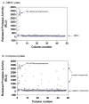
Screen performance in 1536-well plate format. Scatter plot of a DMSO plate (A) and a compound (46 μM) plate (B) in protease release assay in renal mesengial cells. The signal-to-background ratio was 18.2 and the Z′ factor was 0.8 in the DMSO plate, while the signal to background ratio was 16.9 and the Z′ factor was 0.9 in the compound plate.

References

    1. Anderson BM, Noble C., Jr In vitro inhibition of lactate dehydrogenases by kepone. Journal of Agricultural and Food Chemistry. 1977;25:28–31. - PubMed
    1. Blonder E, Klibansky C, DE Vries A. Effects of detergents and choline-containing phospholipids on human spleen glucocerebrosidase. Biochimica et Biophysica Acta. 1976;431:45–53. - PubMed
    1. Boisclar M, Iwata K. HTS using cell-based assays for drug discovery. Am Assoc Cancer Res Educ Book. 2005 April 2005;:363–368.
    1. Corey MJ, Kinders R, Brown L, Vessella R. A very sensitive coupled luminescent assay for cytotoxicity and complement-mediated lysis. Journal of Immunological Methods. 1997;207:43–51. - PubMed
    1. Digan ME, Pou C, Niu H, Zhang J. Evaluation of division-arrested cells for cell-based high throughput screening profiling. Journal of Biomolecular Screening. 2005;10:615–623. - PubMed

Publication types