

Structural and functional biomarkers of prodromal Alzheimer's disease: a high-dimensional pattern classification study
- PMID: 18400519
- PMCID: PMC2682533
- DOI: 10.1016/j.neuroimage.2008.02.043
Structural and functional biomarkers of prodromal Alzheimer's disease: a high-dimensional pattern classification study
Abstract
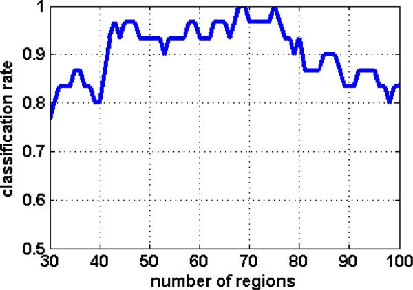
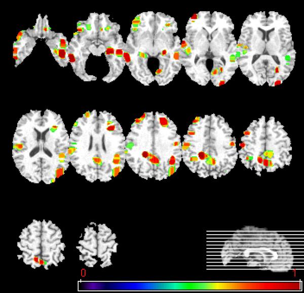
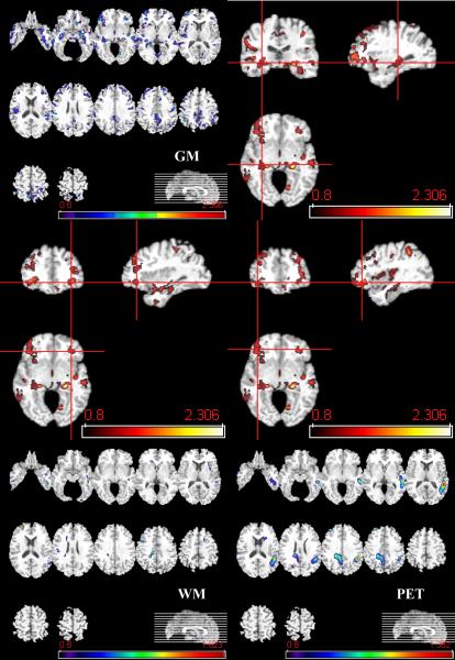
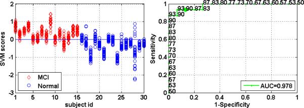
This work builds upon previous studies that reported high sensitivity and specificity in classifying individuals with mild cognitive impairment (MCI), which is often a prodromal phase of Alzheimer's disease (AD), via pattern classification of MRI scans. The current study integrates MRI and PET (15)O water scans from 30 participants in the Baltimore Longitudinal Study of Aging, and tests the hypothesis that joint evaluation of structure and function can yield higher classification accuracy than either alone. Classification rates of up to 100% accuracy were achieved via leave-one-out cross-validation, whereas conservative estimates of generalization performance in new scans, evaluated via bagging cross-validation, yielded an area under the receiver operating characteristic (ROC) curve equal to 0.978 (97.8%), indicating excellent diagnostic accuracy. Spatial maps of regions determined to contribute the most to the classification implicated many temporal, prefrontal, orbitofrontal, and parietal regions. Detecting complex patterns of brain abnormality in early stages of cognitive impairment has pivotal importance for the detection and management of AD.
Figures
References
-
- Ardekani S, Kumar A, Bartzokis G, Sinha U. Exploratory voxel-based analysis of diffusion indices and hemispheric asymmetry in normal aging. Magnetic Resonance Imaging. 2007;25:154–167. - PubMed
-
- Ashburner J, Csernansky JG, Davatzikos C, Fox NC, Frisoni GB, Thompson PM. Computer-assisted imaging to assess brain structure in healthy and diseased brains. The Lancet (Neurology) 2003;2:79–88. - PubMed
-
- Ashburner J, Friston KJ. Voxel-based morphometry: the methods. Neuroimage. 2000;11:805–821. - PubMed
-
- Beresford T, Arciniegas D, Alfers J, Clapp L, Martin B, Beresford H, Du Y, Liu D, Shen D, Davatzikos C, Laudenslager M. Hypercortisolism In Alcohol Dependence and Its Relation to Hippocampal Volume Loss. Journal of Studies on Alcohol. 2006a;67:861–867. - PubMed
-
- Beresford TP, Arciniegas DB, Alfers J, Clapp L, Martin B, Du Y, Liu D, Shen D, Davatzikos C. Hippocampus Volume Loss Due to Chronic Heavy Drinking. Alcoholism: Clinical and Experimental Research. 2006b;30:1866–1870. - PubMed
Publication types
MeSH terms
Grants and funding
LinkOut - more resources
Full Text Sources
Other Literature Sources
Medical
