. 2008 Jun 6;283(23):15694-700.
doi: 10.1074/jbc.M800548200. Epub 2008 Apr 8.

The Hsp60-(p.V98I) mutation associated with hereditary spastic paraplegia SPG13 compromises chaperonin function both in vitro and in vivo

Affiliations

The Hsp60-(p.V98I) mutation associated with hereditary spastic paraplegia SPG13 compromises chaperonin function both in vitro and in vivo

Peter Bross et al. J Biol Chem. .

Abstract

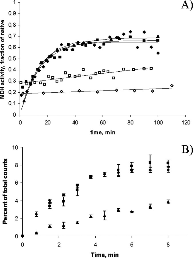
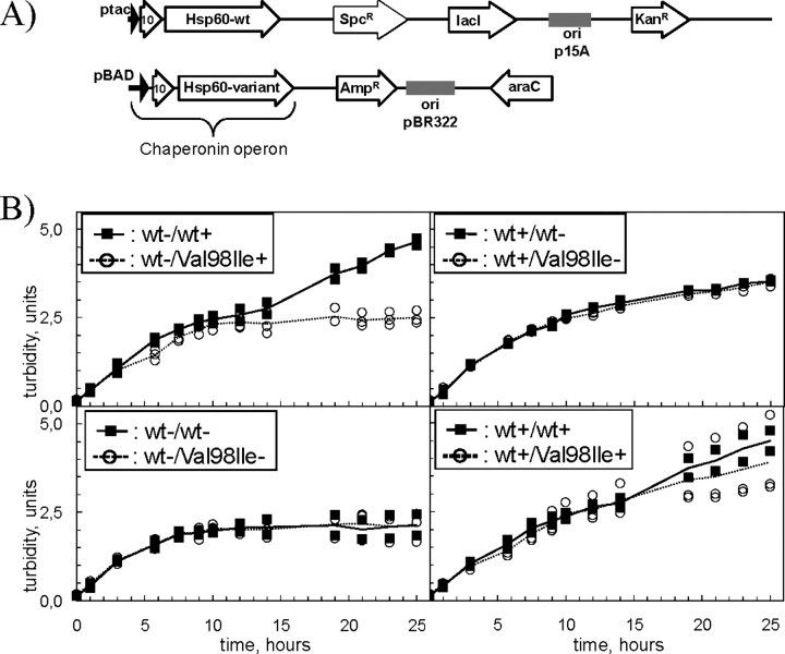
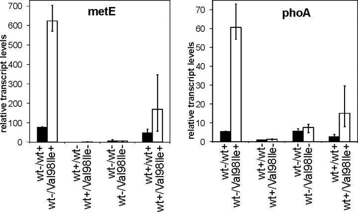
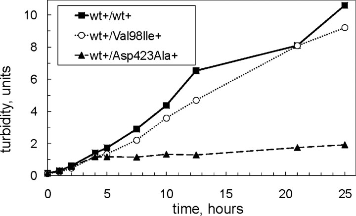
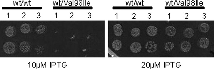
We have previously reported the association of a mutation (c.292G > A/p.V98I) in the human HSPD1 gene that encodes the mitochondrial Hsp60 chaperonin with a dominantly inherited form of hereditary spastic paraplegia. Here, we show that the purified Hsp60-(p.V98I) chaperonin displays decreased ATPase activity and exhibits a strongly reduced capacity to promote folding of denatured malate dehydrogenase in vitro. To test its in vivo functions, we engineered a bacterial model system that lacks the endogenous chaperonin genes and harbors two plasmids carrying differentially inducible operons with human Hsp10 and wild-type Hsp60 or Hsp10 and Hsp60-(p.V98I), respectively. Ten hours after shutdown of the wild-type chaperonin operon and induction of the Hsp60-(p.V98I)/Hsp10 mutant operon, bacterial cell growth was strongly inhibited. No globally increased protein aggregation was observed, and microarray analyses showed that a number of genes involved in metabolic pathways, some of which are essential for robust aerobic growth, were strongly up-regulated in Hsp60-(p.V98I)-expressing bacteria, suggesting that the growth arrest was caused by defective folding of some essential proteins. Co-expression of Hsp60-(p.V98I) and wild-type Hsp60 exerted a dominant negative effect only when the chaperonin genes were expressed at relatively low levels. Based on our in vivo and in vitro data, we propose that the major effect of heterozygosity for the Hsp60-(p.V98I) mutation is a moderately decreased activity of chaperonin complexes composed of mixed wild-type and Hsp60-(p.V98I) mutant subunits.


Figures

FIGURE 1.
FIGURE 1.
A, refolding activity. Refolding of acid-denatured malate dehydrogenase by wild-type Hsp60/Hsp10 (closed diamonds), Hsp60-(p.G67S)/Hsp10 (closed boxes), GroEL/GroES (closed triangles), Hsp60-(p.V98I)/Hsp10 (open boxes), or bovine serum albumin (open diamonds) was measured as described under “Experimental Procedures.” Refolding rates were determined as the slopes of the initial linear part of the curves. B, ATPase activity. ATPase activity of wild-type Hsp60 (diamonds), Hsp60-(p.G67S) (boxes), and Hsp60-(p.V98I) (triangles) in the presence of Hsp10 was determined as described under “Experimental Procedures.” The initial rate for Hsp60-(p.V98I), as determined by the slope of the initial linear part of the curves, was 31% of that of the two wild-type controls. Error bars show the variation in five independent experiments.
FIGURE 2.
FIGURE 2.
A, plasmids used for in vivo studies. E. coli B178 cells deleted for the groESL operon carried two independently replicating plasmids with different selection markers (KanR, kanamycin; AmpR, ampicillin; and SpcR, spectinomycin). Both plasmid types contain an operon with cDNA comprising human Hsp10 and the mature part of wild-type or mutant human Hsp60 under control of the IPTG-inducible tac promoter or the arabinose-inducible BAD promoter. The repressor genes for the tac and BAD promoters, lacI and araC, are also present on the respective plasmids. B, effects of mutant chaperonins on bacterial growth. B178 E. coli cells carrying a deletion of the groESL operon and transformed with different pairs of chaperonin plasmids (see Fig. 2A), were grown at 30 °C in medium supplemented with antibiotics and 0.1 mm IPTG. At an A532 ≈ 0.15 cells were harvested by centrifugation, resuspended in fresh medium, split into four aliquots, the respective inducers were added and growth was monitored for 25 h. The legend denotes induction (+) of the operons with wild-type (wt) or mutant (V98I) Hsp60 on the first/second plasmid or their repression (–). Curves link the average points of three individual transformants of cells carrying Hsp60-(p.V98I) (open circles, stippled line) and two individual transformants carrying wild-type wild-type Hsp60 (closed squares, solid line) on the second plasmid.
FIGURE 3.
FIGURE 3.
Quantitative RT-PCR analysis of metE and phoA transcript levels. RNA purified from samples taken 10 h after inducer shift from the growth experiment shown in Fig. 2A were subjected to qRT-PCR as described under “Experimental Procedures.” metE (left) and phoA (right) transcript levels are given in relation to the endogenous reference gene yeeX and normalized to the average expression levels of cells carrying wild-type Hsp60 on the second plasmid and induced with IPTG only. Columns show the average values obtained from the three cultures harboring Hsp60-(p.V98I) (open columns, V98I) and two cultures harboring wild-type Hsp60 (closed columns, wt) cDNA, respectively, on the second plasmid. The legend denotes induction (+) of the operons with wild-type (wt) or mutant (V98I) Hsp60 on the first/second plasmid or their repression (–). Error bars denote the range of values measured in the biological replicates.
FIGURE 4.
FIGURE 4.
Western blot analysis of MetE and PhoA proteins. Soluble extracts were prepared from samples taken 10 h after inducer shift in the growth experiment shown in Fig. 2B. Aliquots corresponding to 0.5 μgof total soluble protein were subjected to SDS-PAGE followed by Western analysis with anti-MetE or anti-PhoA antibodies. Results from one representative of each cell type are shown. Induction (+) or repression (–) of the operons with wild-type (wt) or mutant (V98I) Hsp60 on the first/second plasmid are indicated below the lower graph.
FIGURE 5.
FIGURE 5.
Assessment of dominant negative effect of mutant co-expression. E. coli B178 cells carrying a deletion of the groESL genes and co-transformed with plasmid pairs carrying chaperonin operons with Hsp10 and wild-type Hsp60 on the first and Hsp10 and Hsp60-(p.V98I), Hsp10-Hsp60-(p.D423A) or wild-type Hsp60, respectively, on the second plasmid were grown up at 30 °C in medium supplemented with 0.1 mm IPTG. At A532 ≈ 0.15 cells were harvested by centrifugation and resuspended in fresh medium supplemented with both arabinose and IPTG. The legend denotes induction (+) or repression (–) of the indicated Hsp60 operons on the first/second plasmid.
FIGURE 6.
FIGURE 6.
Failure of cells co-expressing Hsp60-(pV98I) to form colonies under conditions of limiting Hsp60 levels. The groESL operon was deleted by phage transduction/recombination in E. coli LMG190 cells transformed with the plasmid carrying the IPTG-inducible chaperonin operon with wild-type Hsp60 as described previously (6). One of the selected clones was subsequently transformed with a second plasmid carrying arabinose-inducible chaperonin operons with either wild-type Hsp60 or Hsp60-(p.V98I). Three colonies of each cell type were grown up, and a dilution series was spotted on agar plates containing either 20 μm or 10 μm IPTG as indicated and 0.04% arabinose to partially induce the IPTG/arabinose inducible chaperonin operons. Plates were incubated overnight at 40 °C and photographed.

References

    1. Fink, J. K. (2006) Curr. Neurol. Neurosci. Rep. 6 65–76 - PubMed
    1. Deluca, G. C., Ebers, G. C., and Esiri, M. M. (2004) Neuropathol. Appl. Neurobiol. 30 576–584 - PubMed
    1. Depienne, C., Stevanin, G., Brice, A., and Durr, A. (2007) Curr. Opin. Neurol. 20 674–680 - PubMed
    1. Crosby, A. H., and Proukakis, C. (2002) Am. J. Hum. Genet. 71 1009–1016 - PMC - PubMed
    1. Bross, P., Rugarli, E. I., Casari, G., and Langer, T. (2004) in Mitochondrial Function and Genetics (Koehler, C., and Bauer, M., eds) pp. 97–121, Springer, Heidelberg

Publication types

MeSH terms