. 2008 May 30;283(22):15502-11.
doi: 10.1074/jbc.M708992200. Epub 2008 Apr 8.

Crystal structures of the 70-kDa heat shock proteins in domain disjoining conformation

Affiliations

Crystal structures of the 70-kDa heat shock proteins in domain disjoining conformation

Yi-Wei Chang et al. J Biol Chem. .

Abstract

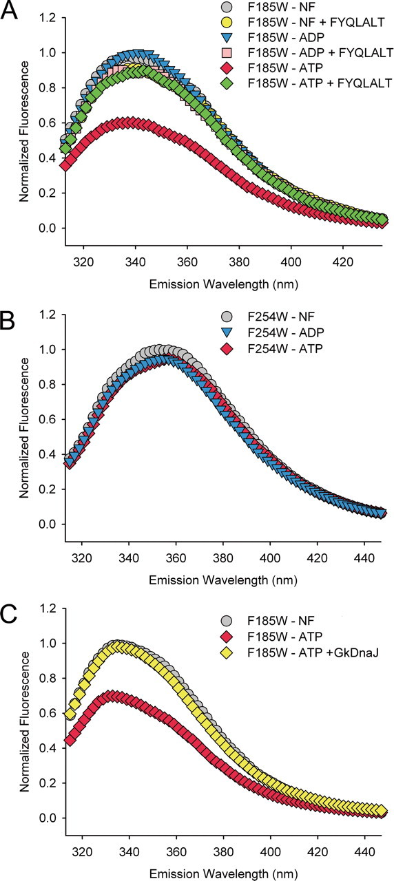
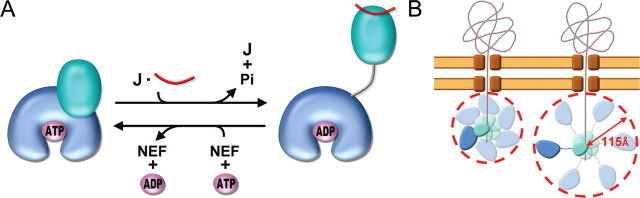
The 70-kDa heat shock proteins (Hsp70s) are highly conserved ATP-dependent molecular chaperones composed of an N-terminal nucleotide binding domain (NBD) and a C-terminal protein substrate binding domain (SBD) in a bilobate structure. Interdomain communication and nucleotide-dependent structural motions are critical for Hsp70 chaperone functions. Our understanding of these functions remains elusive due to insufficient structural information on intact Hsp70s that represent the different states of the chaperone cycle. We report here the crystal structures of DnaK from Geobacillus kaustophilus HTA426 bound with ADP-Mg(2+)-P(i) at 2.37A and the 70-kDa heat shock cognate protein from Rattus norvegicus bound with ADP-P(i) at 3.5A(.) The NBD and SBD in these structures are significantly separated from each other, and they might depict the ADP-bound conformation. Moreover, a Trp reporter was introduced at the potential interface region between NBD and the interdomain linker of GkDnaK to probe environmental changes. Results from fluorescence measurements support the notion that substrate binding enhances the domain-disjoining behavior of Hsp70 chaperones.


Figures

FIGURE 1.
FIGURE 1.
Overall structure of GkDnaKΔC in the ADP-Mg2+-Pi state and intermolecular interactions between symmetry-related GkDnaKΔC molecules. A, ribbon diagram of the GkDnaKΔC-ADP-Mg2+-Pi complex structure and its orthogonal view reveal that the NBD (residues 1-353, blue) and the SBD (residues 365-504, cyan) are connected by an extended linker (residues 354-364, red). The bound nucleotide (ADP, magnesium ion, and Pi) is represented as spheres. B, the extended hydrophobic linker between the NBD and the SBD of GkDnaKΔC (blue) occupies the substrate binding pocket of a neighboring crystallographic symmetry-related GkDnaKΔC molecule (gold).
FIGURE 2.
FIGURE 2.
Peptide binding pocket and adenosine nucleotide binding site of GkDnaKΔC. A, stereo view of the environment surrounding the hydrophobic linker which binds in the substrate binding pocket of a symmetry-related SBD from an adjacent molecule. Direct hydrogen bonds between the two molecules are shown as dashed lines. Models of GkDnaKΔC and its symmetry-related molecule are colored blue and gold, respectively. Interacting residues are represented in stick form. B, stereo view of the adenosine nucleotide binding site in the NBD of GkDnaKΔC. The 2 Fo - Fc electron density map for the bound ADP, magnesium ion, and Pi was contoured at the 1.0 σ level and colored in yellow. Hydrogen bonds and salt bridges between the bound nucleotide and protein are represented as dashed lines. C, schematic representation of the interactions involved in binding the hydrolyzed ATP. Hydrogen bonds and salt bridges between the bound nucleotide and protein are represented as dashed lines with lengths shown in angstroms. Three residues (Arg-314, Ile-315, and Lys-240) that interact hydrophobically with the adenine base of the hydrolyzed ATP are boxed.
FIGURE 3.
FIGURE 3.
Comparison of ADP-Mg2+-Pi bound and nucleotide-free Hsp70 structures. A, structural superposition between the NBD of GkDnaKΔCinthe ADP-Mg2+-Pi state (blue) and that of EcoDnaK in nucleotide-free state (yellow). The most structurally disparate region between GkDnaKΔC and EcoDnaK is colored in red. The bound nucleotide in the GkDnaKΔC NBD is shown in the stick form. The arrows indicate the possible closing behavior of the pocket upon nucleotide binding. B, diagram representation of the large-scale spatial difference for domain arrangement between GkDnaKΔC (blue) and bHsc70ΔC (orange). The NBDs of two structures are superimposed. Helixes A in both structures are colored red. C, stereo view of a structural superimposition of the ADP-Mg2+-Pi state GkDnaKΔC (blue) with the nucleotide-free state bHsc70ΔC (NBD, orange; SBD, yellow). Residues in the NBD of bHsc70ΔC that participate in domain-domain interactions are highly conserved in the GkDnaKΔC NBD. Equivalent residues in the bHsc70ΔC SBD are also highly conserved in the GkDnaKΔC SBD (bHsc70:GkDnaK, Arg-416:Arg-383, Asn-417:Asn-384, Glu-513:Glu-479, Ile-515:Ile-481, Val-519:Ile-485, Ala-522:Ala-488). D, diagram representation of the large-scale spatial difference for domain arrangement between GkDnaKΔC (blue) and yeast Sse1 (magenta). The NBDs of these two structures are superimposed. E, structural superimposition of the NBD and interdomain linker from the GkDnaKΔC (blue) and the yeast Sse1 (magenta). The interdomain linkers from both structure are colored yellow. Residue Phe-185 of GkDnaKΔC is represented as a spheres and colored red.
FIGURE 4.
FIGURE 4.
Examination of the nucleotide- and substrate-induced conformational changes in the GkDnaKΔC. A, the normalized fluorescence emission spectra of GkDnaKΔC-F185W in the nucleotide-free state (NF, gray), ADP-bound state (blue), ATP-bound state (red), nucleotide-free state with substrate FYQLALT (yellow), ADP-bound state with substrate FYQLALT (pink), and ATP-bound state with substrate FYQLALT (green). B, the normalized fluorescence emission spectra of GkDnaKΔC-F254W mutant in different nucleotide binding states. C, the GkDnaJ-induced effect (yellow) on the reversal of the ATP-induced fluorescence quenching for the GkDnaKΔC-F185W mutant.
FIGURE 5.
FIGURE 5.
Overall structure, adenosine nucleotide binding site, and the highly flexible SBD of rHsc70ΔC in the ADP-Pi state. A, ribbon drawing of the rHsc70ΔC-ADP-Pi complex. The NBD (residues 2-382) is colored green, and the bound nucleotide (ADP and Pi) are represented as spheres. The SBD (residues 383-543) and hydrophobic linker region are unassignable here. B, the crystals of rHsc70ΔC were analyzed by SDS-PAGE and visualized by dye-staining. Lane 1, the rHsc70ΔC solution used in the crystallization reaction. Lane 2, the rHsc70ΔC crystals. C, the superimposition of ADP-Mg2+-Pi-bound form of GkDnaKΔC (blue) and nucleotide-free form of bHsc70ΔC (orange) on the crystal packing of the ADP-Pi-bound form of rHsc70ΔC (gray). The plausible space for the SBD of rHsc70ΔCis encircled by magenta dashed lines. D, stereo view of the environment surrounding the adenosine nucleotide binding site in the NBD of rHsc70ΔC. The 2 Fo - Fc electron density map for the bound ADP-Pi was contoured at 1.0 σ (gray) and 3.5 σ (yellow) levels. The stick models indicate residues interacting with ADP or Pi. Dashed lines indicate hydrogen bonds and salt bridges between the bound nucleotide and protein. E, the superimposition of bound adenosine nucleotides among GkDnaKΔC-ADP-Mg2+-Pi, rHsc70ΔC-ADP-Pi, and bovine NBD (K71M)-ATP-Mg2+-K+ precatalytic state structures. All atoms are represented by sticks except Mg2+, which is represented by spheres. Coloring has the following pattern: carbon (gray), nitrogen (cyan), oxygen (red), phosphorous and Mg2+ of GkDnaKΔC (blue), phosphorous and Mg2+ of bovine NBD (K71M) (green), phosphorous of rHsc70ΔC (yellow), and Asp-199 of rHsc70ΔC (light yellow) and its corresponding residues in GkDnaKΔC (light blue) and bovine NBD (K71M) (light green). The β- and γ-phosphate of pre- of post-hydrolyzed ATP are linked by dashed lines with lengths shown in angstroms.
FIGURE 6.
FIGURE 6.
Implication of Hsp70 chaperone cycle and benefits for domain-disjoining conformation of Hsp70s. A, schematic representation for the domain rearrangement of Hsp70. The NBD, SBD, and linker of Hsp70 are colored blue, cyan, and gray, respectively. The cochaperones J-domain ATPase activating protein and nucleotide exchange factor are symbolized by letters J and NEF, respectively. The substrate of Hsp70 is represented as red ribbons, and ATP and ADP are as magenta ellipsoids. B, schematic diagram depicting how luminal Hsp70s enlarge their excluded-volume effect by extending the two-lobed conformation (left, before; right, after) while conducting the post-translational translocation of precursor proteins across membranes. The two-lobed structures are Hsp70 molecules in which the NBDs are colored blue, the SBDs are colored cyan, and the interdomain linkers are colored gray. The brown structures represent the channel complexes that span two-layer membranes, and ribbons represent precursor proteins being imported into the lumen. The possible distributing area of the substrate-bound Hsp70 are represented by paler versions of Hsp70 molecules and circled by red dashed lines.

References

    1. Young, J. C., Agashe, V. R., Siegers, K., and Hartl, F. U. (2004) Nat. Rev. Mol. Cell Biol. 5 781-791 - PubMed
    1. Bukau, B., and Horwich, A. L. (1998) Cell 92 351-366 - PubMed
    1. Hartl, F. U. (1996) Nature 381 571-579 - PubMed
    1. Neupert, W., and Brunner, M. (2002) Nat. Rev. Mol. Cell Biol. 3 555-565 - PubMed
    1. Wegele, H., Muller, L., and Buchner, J. (2004) Rev. Physiol. Biochem. Pharmacol. 151 1-44 - PubMed

Publication types

Substances