Review
. 2008 Apr;21(2):305-33, table of contents.
doi: 10.1128/CMR.00060-07.

Cavitary pulmonary disease

Affiliations
Review

Cavitary pulmonary disease

L Beth Gadkowski et al. Clin Microbiol Rev. 2008 Apr.

Abstract

A pulmonary cavity is a gas-filled area of the lung in the center of a nodule or area of consolidation and may be clinically observed by use of plain chest radiography or computed tomography. Cavities are present in a wide variety of infectious and noninfectious processes. This review discusses the differential diagnosis of pathological processes associated with lung cavities, focusing on infections associated with lung cavities. The goal is to provide the clinician and clinical microbiologist with an overview of the diseases most commonly associated with lung cavities, with attention to the epidemiology and clinical characteristics of the host.


Figures

FIG. 1.
FIG. 1.
Sequelae of severe Staphylococcus aureus pneumonia in a patient with multiple other comorbidities. The left panel illustrates the plain chest radiographic appearance, with multiple areas of fibrosis and a residual cavity in the medial right upper lobe. The right panel shows the same cavity using computed tomography.
FIG. 2.
FIG. 2.
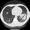
Left lower lobe cavitary pneumonia due to Rhodococcus equi with concurrent bacteremia in a patient with advanced human immunodeficiency virus.
FIG. 3.
FIG. 3.
Extensive cavitary lung disease due to Mycobacterium tuberculosis visualized by plain chest radiography (left) and computed tomography (right). Note the typical upper lobe predominance and extensive fibronodular infiltrates.
FIG. 4.
FIG. 4.
Mycobacterium avium complex of the fibrocavitary type in a 52-year-old woman with chronic obstructive pulmonary disease visualized by plain radiography (left) and computed tomography (right).
FIG. 5.
FIG. 5.
Mycobacterium kansasii pulmonary disease presenting as 8 months of hemoptysis without any systemic symptoms in a 16-year-old girl. The thin-walled cavities (in the right apex) are relatively subtle on plain chest radiography (left) but are readily apparent by computed tomography, as is the accompanying bronchiectasis (right).
FIG. 6.
FIG. 6.
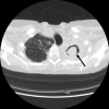
Aspergilloma (round mass in the left upper lobe) visualized by computed tomography in a young man. The etiology of the underlying cavity was unknown in this case.
FIG. 7.
FIG. 7.
Invasive aspergillosis in a 52-year-old man with systemic lupus erythematosus who had been on chronic high-dose corticosteroids and azathioprine. The plain chest radiograph (left) is suggestive of septic pulmonary emboli, with multiple large nodules bilaterally, at least two of which contain central cavities. The right panel demonstrates one of these thick-walled cavities as seen by computed tomography. This patient also had Aspergillus in the brain, which is a common site of metastatic spread in immunocompromised hosts.

References

    1. Abehsera, M., D. Valeyre, P. Grenier, H. Jaillet, J. P. Battesti, and M. W. Brauner. 2000. Sarcoidosis with pulmonary fibrosis: CT patterns and correlation with pulmonary function. Am. J. Roentgenol. 174:1751-1757. - PubMed
    1. Aberg, J. A., L. M. Mundy, and W. G. Powderly. 1999. Pulmonary cryptococcosis in patients without HIV infection. Chest 115:734-740. - PubMed
    1. Aberle, D. R., G. Gamsu, and D. Lynch. 1990. Thoracic manifestations of Wegener granulomatosis: diagnosis and course. Radiology 174:703-709. - PubMed
    1. Abramson, S. 2001. The air crescent sign. Radiology 218:230-232. - PubMed
    1. Aguado, J. M., G. Obeso, J. J. Cabanillas, M. Fernandez-Guerrero, and J. Ales. 1990. Pleuropulmonary infections due to nontyphoid strains of Salmonella. Arch. Intern. Med. 150:54-56. - PubMed

Publication types