. 2008 Jun;82(12):5879-86.
doi: 10.1128/JVI.02432-07. Epub 2008 Apr 9.

A subgenomic segment of Theiler's murine encephalomyelitis virus RNA causes demyelination

Affiliations

A subgenomic segment of Theiler's murine encephalomyelitis virus RNA causes demyelination

Gleb Baida et al. J Virol. 2008 Jun.

Abstract

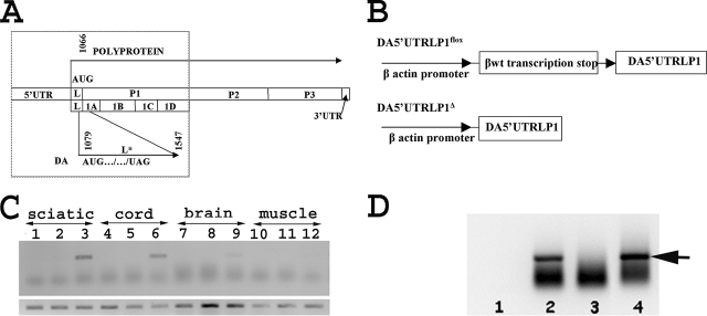
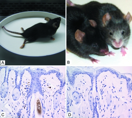
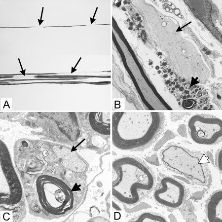
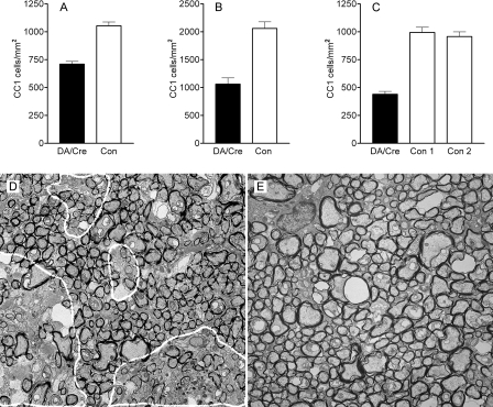
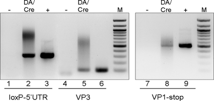
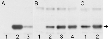
The DA strain of Theiler's murine encephalomyelitis virus (TMEV) causes a persistent central nervous system (CNS) infection of mice with a restricted virus gene expression and induces an inflammatory demyelinating disease that is thought to be immune mediated and a model of multiple sclerosis (MS). The relative contribution of virus vis-à-vis the immune system in the pathogenesis of DA-induced white matter disease remains unclear, as is also true in MS. To clarify the pathogenesis of DA-induced demyelination, we used Cre/loxP technology to generate a transgenic mouse that has tamoxifen (Tm)-inducible expression of a subgenomic segment of DA RNA in oligodendrocytes and Schwann cells. Tm-treated young transgenic mice developed progressive weakness leading to death, with abnormalities of oligodendrocytes and Schwann cells and demyelination, but without inflammation, demonstrating that DA virus can play a direct pathogenic role in demyelination. Tm treatment of mice at a later age resulted in milder disease, with evidence of peripheral nerve remyelination and focal fur depigmentation; surviving weak mice had persistent expression of the recombined transgene in the CNS, suggesting that the DA subgenomic segment can cause cellular dysfunction but not death, possibly similar to the situation seen during DA virus persistence. These studies demonstrate that DA RNA or a DA protein(s) is toxic to myelin-synthesizing cells. This Cre/loxP transgenic system allows for spatially and temporally controlled expression of the viral transgene and is valuable for clarifying nonimmune (and immune) mechanisms of demyelination induced by TMEV as well as other viruses.


Figures

FIG. 1.
FIG. 1.
The DA virus genome, subgenomic transgene, and amplified products following PCR and RT-PCR of tissues. (A) The TMEV single-stranded RNA genome of approximately 8,100 nt (not drawn to scale). The 5′UTR precedes the polyprotein coding region, which has the leader (L) coding region at its most amino terminus. The genomic region of P1 is divided into 1A, 1B, 1C, and 1D coding regions, which synthesize the structural capsid proteins VP4, VP2, VP3, and VP1, respectively. The polyprotein's initiation codon is at nt 1066, while the L* initiation codon in the case of the DA strain is at nt 1079. There is a stop codon (UAG) for L* at nt 1547. The dotted line shows the subgenomic region that is included in the transgene. (B) Schematic representation (not drawn to scale) of DA5′UTRLL*P1 (top panel), showing the loxP sites (that flank an upstream transcription stop) as triangles, and DA5′UTRLL*P1Δ (bottom panel) after Cre-induced recombination. (C) The upper panel shows an agarose gel of amplified products following PCR with primers to detect recombination of genomic DNA from homogenates of sciatic nerve (lanes 1 to 3), spinal cord (lanes 4 to 6), brain (lanes 7 to 9), and muscle (lanes 10 to 12) from the following animals: a Tm-treated mouse that only carries the DA subgenomic transgene (lanes 1, 4, 7, and 10); a DA/Cre mouse not treated with Tm (lanes 2, 5, 8, and 11); a DA/Cre mouse treated with Tm (lanes 3, 6, 9, and 12). Only neural tissues from Tm-treated DA/Cre mice showed an amplified band of the predicted size of ∼300 bp. The lower panel shows amplified products that were obtained from a separate PCR using the primer pair that detects the DA subgenomic transgene irrespective of recombination. The bands at the predicted size of 200 bp are present in all lanes. (D) Agarose gel of amplified products following RT-PCR, using primers to detect recombination, from RNA extracted from the spinal cord (lane 2), lung (lane 3), and brain (lane 4) from a Tm-treated DA/Cre mouse. Lane 1 has no cDNA template. A band (arrow) of the predicted size of ∼300 bp was present in the spinal cord and brain, but not lung.
FIG. 2.
FIG. 2.
Phenotype of DA/Cre mice. (A) A DA/Cre mouse 22 days after Tm treatment (begun at 21 days of age), showing paralysis of the hind legs and weakness of the forelegs. (B) A DA/Cre mouse 3 months after Tm treatment (begun at 7 weeks of age) with a littermate control. The DA/Cre mouse shows depigmentation of fur in several regions. (C) Hematoxylin staining of perioral skin from the littermate control shown in panel B, demonstrating brown melanin pigment in dermal dendritic melanocytes, follicular bulb melanocytes, and hair shaft. Magnification, ×400. (D) Hematoxylin staining of depigmented perioral skin of the DA/Cre mouse shown in panel B, demonstrating no melanin pigment. Magnification, ×400.
FIG. 3.
FIG. 3.
Pathology of Tm-treated DA/Cre mice. (A to C) DA/Cre mouse euthanized 18 days after Tm treatment (begun at 4 weeks of age). (D) DA/Cre mouse euthanized 3 months after Tm treatment (begun at 7 weeks of age). Arrows, some demyelinated axons; closed arrowhead, myelin debris; open arrowhead, remyelinated axon. (A) Teased fiber preparation from the sciatic nerve showing a single osmicated myelinated axon, demonstrating two foci of segmental demyelination (upper panel) and a bundle of adjacent myelinated axons demonstrating multiple foci of segmental demyelination (lower panel). (B) Electron micrograph of longitudinally oriented sciatic nerve, demonstrating an intact demyelinated axon. A Schwann cell adjacent to the demyelinated axon contains myelin debris. Magnification, ×3,000. (C) Electron micrograph of transversely oriented sciatic nerve, demonstrating intact demyelinated axon surrounded by Schwann cell cytoplasm with degenerative changes and myelin debris. Magnification, ×5,400. (D) Transverse section through the sciatic nerve, demonstrating a thin myelin sheath, characteristic of remyelination, around a large axon. The adjacent myelin sheaths are of the appropriate thickness for the diameter of the axons they surround. Magnification, ×5,400.
FIG. 4.
FIG. 4.
Abnormalities of the CNS white matter of Tm-treated DA/Cre mice. Nonoverlapping rectangular areas (∼50,000 μm2 each) of white matter were analyzed for CC1-positive cells. (A) The mean number of CC1 antibody staining cells from 20 areas of 14 separate sections of the spinal cord from three DA/Cre mice (that were treated with Tm for 10 days starting at ∼3 weeks of age and euthanized when moribund ∼1.5 to 3 weeks after the last Tm dose) was significantly less (P < 0.001) than in 22 areas of 9 sections from the spinal cord of three littermates that carried no or a single transgene and had identical Tm treatment. (B) The mean number of CC1 antibody staining cells from six areas of three sections of the corpus callosum from one of the DA/Cre transgenic mice that were immunostained in panel A was significantly decreased (P < 0.001) compared to four areas from two sections of one of the nontransgenic littermates that were immunostained in panel A. (C) The mean number of CC1 antibody staining cells from seven areas from six sections of the optic nerve from a DA/Cre mouse that was treated with Tm for 10 days starting at ∼4 weeks of age and euthanized when moribund 19 days later was significantly less (P < 0.001) than 12 areas from six sections of the optic nerve from two single transgenic littermates that had identical Tm treatment. (D) Electron micrograph of the optic nerve of the DA/Cre mouse (that was immunostained in panel C), showing clusters of unmyelinated axons in the optic nerve (circled areas). Magnification, ×2,500. (E) Electron micrograph of the optic nerve of a control Tm-treated littermate that was immunostained in panel C. Magnification, ×2,500.
FIG. 5.
FIG. 5.
The recombined DA subgenomic construct is present and expressed in a surviving DA/Cre mouse ∼3 months after Tm treatment. RT-PCR of CNS from a DA/Cre mouse 3 months after Tm treatment that was begun at age 7 weeks. A series of primers was used that covered the full length of the transgene's RNA from the 5′ end (LoxP-5′UTR; lanes 1 to 3) through some of the coding region (VP3; lanes 4 to 6) to the termination codon (VP1-stop; lanes 7 to 9). The sizes of the amplified segment correspond to the expressed full length of the recombined transgene. -, no RT; +, plasmid; M, marker.
FIG. 6.
FIG. 6.
Western immunoblot results, showing a decrease in expression of GFP when DA L protein is expressed. All transfections involved an identical amount of a GFP expression vector, peGFP-N1, and an identical total amount of DNA. The arrow shows the predicted mobility of GFP. Homogenates of BHK-21 cells were subjected to sodium dodecyl sulfate-polyacrylamide gel electrophoresis followed by Western blot analysis using anti-GFP antibody and then ECL-Plus Western blot detection. (A) Results for homogenate not transfected (lane 1) or transfected with peGFP-N1 (lane 2) along with pcDNA3.1 vector or peGFP-N1 and the DA L coding region cloned into peGFPN-1 (so that GFP is fused to its carboxyl end) (lane 3). This Western blot is representative and shows a decrease in the expression of GFP as a result of cotransfection with L-GFP. Longer exposures showed that there was also minimal expression of L-GFP, indicating that its expression was also decreased. (B) A separate Western blot showing homogenate not transfected (lane 1) or transfected with peGFP-N1 along with the recombined DA5′UTRLL*P1 transgene (lane 2), with pcDNA3.1 vector (lane 3), or with a bicistronic luciferase vector with the DA IRES in the intercistronic region (lane 4). The Western blot is representative and shows a decrease in the expression of GFP when cotransfected with the recombined transgene but not with a bicistronic vector containing the DA IRES. (C) A separate Western blot of homogenates of BHK-21 cells showing transfection of peGFP-N1 along with the recombined transgene (lane 1) or the unrecombined transgene (lane 2). This Western blot shows that there is a decrease in GFP expression following cotransfection with the recombined, but not the unrecombined, transgene.

Similar articles

Cited by

References

    1. Chen, H. H., W. P. Kong, L. Zhang, P. L. Ward, and R. P. Roos. 1995. A picornaviral protein synthesized out of frame with the polyprotein plays a key role in a virus-induced immune-mediated demyelinating disease. Nat. Med. 1927-931. - PubMed
    1. Doerflinger, N. H., W. B. Macklin, and B. Popko. 2003. Inducible site-specific recombination in myelinating cells. Genesis 3563-72. - PubMed
    1. Drescher, K. M., and S. M. Tracy. 2007. Injection of the sciatic nerve with TMEV: a new model for peripheral nerve demyelination. Virology 359233-242. - PMC - PubMed
    1. Dye, M. J., and N. J. Proudfoot. 2001. Multiple transcript cleavage precedes polymerase release in termination by RNA polymerase II. Cell 105669-681. - PubMed
    1. Dye, M. J., and N. J. Proudfoot. 1999. Terminal exon definition occurs cotranscriptionally and promotes termination of RNA polymerase II. Mol. Cell 3371-378. - PubMed

Publication types

MeSH terms

LinkOut - more resources