. 2008 Jun;82(12):6045-51.
doi: 10.1128/JVI.02392-07. Epub 2008 Apr 9.

Site 1 protease is required for proteolytic processing of the glycoproteins of the South American hemorrhagic fever viruses Junin, Machupo, and Guanarito

Affiliations

Site 1 protease is required for proteolytic processing of the glycoproteins of the South American hemorrhagic fever viruses Junin, Machupo, and Guanarito

Jillian M Rojek et al. J Virol. 2008 Jun.

Abstract

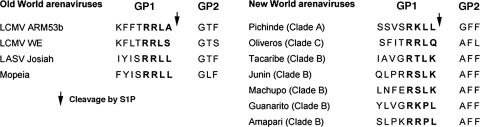
The cellular proprotein convertase site 1 protease (S1P) has been implicated in the proteolytic processing of the glycoproteins (GPs) of Old World arenaviruses. Here we report that S1P is also involved in the processing of the GPs of the genetically more-distant South American hemorrhagic fever viruses Guanarito, Machupo, and Junin. Efficient cleavage of Guanarito virus GP, whose protease recognition sites deviate from the reported S1P consensus sequence, indicates a broader specificity of S1P than anticipated. Lack of GP processing of Junin virus dramatically reduced production of infectious virus and prevented cell-to-cell propagation. Infection of S1P-deficient cells resulted in viral persistence over several weeks without the emergence of escape variants able to use other cellular proteases for GP processing.


Figures

FIG. 1.
FIG. 1.
Amino acid sequences at the sites of proteolytic processing of the GP precursor (GPC) into GP1 and GP2 for selected Old World and New World arenaviruses. The putative recognition sequences of S1P protease are indicated in bold, and the site of S1P cleavage is marked with an arrow.
FIG. 2.
FIG. 2.
Expression and function of flag-tagged arenavirus GPs. (A) Schematic representation of flag-tagged arenavirus GPs: gray boxes and boxes labeled SSP correspond, respectively, to the transmembrane domain and stable signal peptide. Arrows indicate sites of proteolytic processing. (B) The expression of C-terminally flag-tagged GPs of AMPV, GTOV, JUNV, MACV, and LASV as well as a green fluorescent protein (GFP) control in lysates of transfected HEK293T cells was examined by Western blotting using an antiflag antibody. The positions of the fully glycosylated (GPC) and underglycosylated (GPCu) GPC species and mature GP2 are indicated. (C) Generation of recombinant retroviral vectors pseudotyped with arenavirus GPs. The packaging cell line GP2293 stably transfected with murine leukemia virus (MLV) gag and pol was cotransfected with a plasmid containing the packable MLV genomic plasmid pLZRS-Luc-gfp, carrying a luciferase and a GFP reporter (22), and an expression plasmid for the heterologous recombinant GP. Retroviral pseudotypes are released into the cell supernatant. LTR, long terminal repeat. (D) Infection of cells with retroviral pseudotypes. Retroviral pseudotypes were generated with wild-type and flag-tagged GPs of AMPV, GTOV, JUNV, MACV, and LASV and, as a control, GFP. Retroviral pseudotype-containing supernatants were added to Vero E6 cells, and 48 h later, infection was assessed by a Steady Glo luciferase assay (n = 3; error bars indicate standard deviations).
FIG. 3.
FIG. 3.
S1P is required for processing and function, but not trafficking, of the GPs of human-pathogenic arenaviruses. (A) Expression of arenavirus GPs in S1P-deficient cells. S1P-deficient SRD12B cells and wild-type CHOK1 cells were transfected with flag-tagged GPs of AMPV, GTOV, JUNV, MACV, and LASV or with GFP as a control, and 48 h later, cell lysates were analyzed by Western blotting with an antiflag antibody. (B) Complementation of SRD12B cells with recombinant S1P. SRD12B cells were cotransfected with the indicated flag-tagged GPs and either empty control vector (SRD12B cells) or an expression plasmid for S1P (SRD12B cells + S1P). Viral GPs were detected by Western blotting as described for panel A. (C) S1P processing is not required for cell surface expression of arenaviral GPs. SRD12B cells and CHOK1 cells were transfected with the indicated GPs or, as a control, an empty plasmid (mock). After 48 h, cells were detached by nonenzymatic treatment and live, nonpermeabilized cells were stained with MAb 83.6 to AMPV GP2 and LASV GP2 (21) and MAb BE08 to JUNV GP1 (16). Primary antibody was detected with a phycoerythrin (PE)-labeled secondary antibody and analyzed by flow cytometry using a FACSCalibur flow cytometer (13). Data were acquired and analyzed using Cell Quest and FloJo software packages. In dot plots, the y axis represents forward scatter and the x axis represents PE fluorescence intensity. (D) S1P-mediated processing is required for the function of arenavirus GPs. SRD12B cells and CHOK1 cells were cotransfected with a plasmid expressing MLV gag and pol, the MLV genomic plasmid pLZRS-Luc-gfp, and expression plasmids for the indicated GPs, and 48 h later, conditioned supernatants were harvested, cleared, and added to Vero E6 monolayers. Infection was detected by a luciferase assay as described in the legend for Fig. 2D. Note that the y axis has a log scale.
FIG. 4.
FIG. 4.
Mutagenesis of the putative S1P cleavage site of JUNV GP. (A) Sequence of wild-type and mutant JUNV GPs at the putative S1P cleavage site (arrow) with mutated amino acids show in bold. The sequence of MACV is shown for comparison. (B) Wild-type and mutant JUNV flag-tagged GPs were expressed in HEK293T cells, and total protein lysates were analyzed by Western blotting as described for Fig. 2B. The positions of GPC, the putative underglycosylated GPC form GPCu, and mature GP2 are indicated. (C) Densitometric analysis of the blot in panel B. Densitometry was performed as described previously (6), and the ratios of the signal intensities for GP2 (IGP2) to GPC (IGPC) were calculated (GP2/GPC) for wild-type and mutant JUNV GP-flag.
FIG. 5.
FIG. 5.
S1P processing is required for the production of infectious JUNV and cell-to-cell propagation. (A) Infection of SRD12B and CHOK1 cells with JUNV Candid 1. Cells (96-well plates) were infected with 100 PFU of JUNV Candid 1. At the indicated times, cells were fixed and stained with MAb BG12 for JUNV NP. The number of infectious foci was determined in each well (n = 3; error bars indicate standard deviations [SD]). (B) Representative infectious foci in SDR12B and CHOK1 cells at 48 h postinfection. Bar = 20 μm. (C) Production of infectious JUNV in SRD12B and CHOK1 cells. Cells were infected with JUNV Candid 1 at an MOI of 1. Cell supernatants were harvested at the indicated times, and infectious virus titers were determined by a plaque assay on Vero E6 cells. A compilation of three independent experiments is shown (means ± SD). (D to F) Analysis of the composition of JUNV particles produced in SRD12B and CHOK1 cells. Triplicate samples of 5 × 10⁶ SRD12B (SRD) and CHOK1 (CHO) cells grown in T75 flasks were infected with JUNV Candid 1 at an MOI of 1. (D) Cell culture supernatants were harvested after 48 h, and infectious virus titers were determined by a plaque assay on Vero E6 cells. (E) Detection of viral proteins by Western blotting. Supernatants were subjected to ultracentrifugation through a 20% sucrose cushion (6). Pellets were solubilized in hot SDS-PAGE loading buffer, and viral proteins were separated by SDS-PAGE under nonreducing conditions. Blots were probed with MAbs GB03-BE08 (anti-JUNV GP1) and SA02-BG12 (anti-JUNV NP) (16) combined with a biotinylated secondary antibody and horseradish peroxidase-conjugated streptavidin. Signals were detected using the Super Signal West Femto chemiluminescence detection kit from Pierce. (F) Densitometric analysis of the blots in panel E. Densitometry was performed as described in the legend to Fig. 4C, and the ratios of the signal intensities for GP1 (IGP1) to NP (INP) were calculated (GP1/NP1) for virion particles from SRD12B cells (SRD) and CHOK1 cells (CHO). (G) Persistent infection of SRD12B cells with JUNV Candid 1 does not result in escape variants. SRD12B (three independent cell cultures) and CHOK1 cells were infected with JUNV Candid 1 at an MOI of 1 and passaged every 3 days. At the indicated times, virus titers were determined by a plaque assay. The results of one representative example out of three independent experiments is shown.

References

    1. Beyer, W. R., D. Popplau, W. Garten, D. von Laer, and O. Lenz. 2003. Endoproteolytic processing of the lymphocytic choriomeningitis virus glycoprotein by the subtilase SKI-1/S1P. J. Virol. 772866-2872. - PMC - PubMed
    1. Brown, M. S., and J. L. Goldstein. 1997. The SREBP pathway: regulation of cholesterol metabolism by proteolysis of a membrane-bound transcription factor. Cell 89331-340. - PubMed
    1. Buchmeier, M. J., J. C. de la Torre, and C. J. Peters. 2007. Arenaviridae: the viruses and their replication, p. 1791-1828. In D. L. Knipe and P. M. Howley (ed.), Fields virology, 4th ed. Lippincott-Raven, Philadelphia, PA.
    1. Enria, D. A., and J. G. Barrera Oro. 2002. Junin virus vaccines. Curr. Top. Microbiol. Immunol. 263239-261. - PubMed
    1. Geisbert, T. W., and P. B. Jahrling. 2004. Exotic emerging viral diseases: progress and challenges. Nat. Med. 10S110-S1121. - PubMed

Publication types

MeSH terms

LinkOut - more resources