

doi: 10.1128/JVI.00074-08.
Epub 2008 Apr 9.
Cleavage of group 1 coronavirus spike proteins: how furin cleavage is traded off against heparan sulfate binding upon cell culture adaptation
Affiliations
- PMID: 18400867
- PMCID: PMC2395124
- DOI: 10.1128/JVI.00074-08
Item in Clipboard
Cleavage of group 1 coronavirus spike proteins: how furin cleavage is traded off against heparan sulfate binding upon cell culture adaptation
J Virol.
2008 Jun.
Abstract
A longstanding enigmatic feature of the group 1 coronaviruses is the uncleaved phenotype of their spike protein, an exceptional property among class I fusion proteins. Here, however, we show that some group 1 coronavirus spike proteins carry a furin enzyme recognition motif and can actually be cleaved, as demonstrated for a feline coronavirus. Interestingly, this feature can be lost during cell culture adaptation by a single mutation in the cleavage motif; this, however, preserves a heparan sulfate binding motif and renders infection by the virus heparan sulfate dependent. We identified a similar cell culture adaptation for the human coronavirus OC43.
Figures
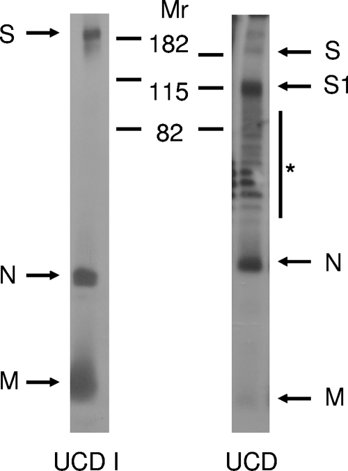
FCoV UCD virions, but not FCoV UCD1 virions, contain cleaved spike proteins. Radiolabeled FCoV UCD1 virions were immunoprecipitated from infected cell culture supernatant using an FCoV RM antiserum and subjected to SDS-PAGE. FCoV UCD virions were subjected to SDS-PAGE, followed by Western blotting using the same antiserum to visualize FCoV-specific proteins. The positions of the molecular weight markers (Mr; in thousands) are indicated. Arrows indicate the FCoV-specific S, S1, N, and M proteins. The asterisk indicates a ladder of nonspecific bands that was consistently observed with feces-derived viral samples.
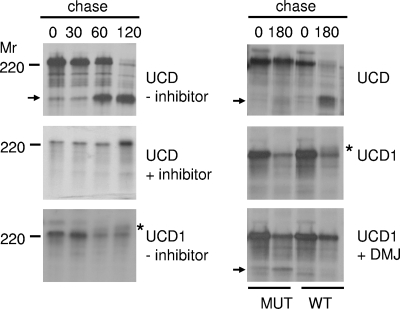
The spike protein of FCoV UCD, but not that of FCoV UCD1, is cleaved by a furin-like protease. OST7-1 cells infected with vaccinia virus vTF7-3 were transfected with plasmids carrying either the UCD or UCD1 spike gene under the control of bacteriophage T7 transcription regulatory elements. The cells were pulse-labeled with 35S-amino acids for 1 h and subsequently chased for 0, 30, 60, 120, or 180 min. Where indicated by a plus, the furin inhibitor dec-RVKR-cmk (75 μM) (left panel) or the mannosidase I inhibitor DMJ (1 mM) (right panel) was added to the culture media and kept present throughout the experiment. Spike cleavages of the wild-type (WT) and mutant (MUT) UCD and UCD1 proteins were also compared (see text for details). The cells were lysed and processed for immunoprecipitation using FCoV RM antiserum. The immunoprecipitates were analyzed by SDS-PAGE. The position of a molecular weight marker (Mr) is shown on the left side of the panels. The asterisks indicate the positions of the mature UCD1 spike protein. The arrows indicate the positions of the S1 cleavage products. Only the relevant portion of the gel is shown.
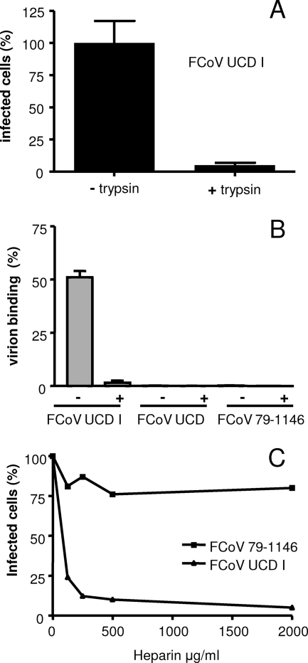
Heparin binds to FCoV UCD1 and inhibits its infectivity. (A) Sucrose gradient-purified FCoV UCD1 particles were treated with TPCK (tosylsulfonyl phenylalanyl chloromethyl ketone)-trypsin (25 μg/ml) for 30 min at room temperature (+trypsin) or mock treated (−trypsin) prior to the inoculation of FCWF cells. The cells were fixed at 18 h postinfection. The number of infected cells relative to the number of cells infected after inoculation with mock-treated viruses was determined by immunoperoxidase staining using the FCoV RM antiserum. (B) Similar quantities of FCoV UCD1, UCD, and 79-1146 virions (as determined by quantitative TaqMan RT-PCR [19]) were incubated for 1 h at 4°C with heparin-agarose beads in the presence (+) or absence (−) of heparin (500 ng/ml; added 1 h before addition of the beads). After three washes, the amounts of virions adsorbed to the beads, expressed again as viral genome equivalents, were determined relative to the amount of input RNA. (C) FCoV UCD1 and 79-1146 were incubated with different concentrations of heparin for 1 h at 4°C prior to the inoculation of FCWF cells. The cells were fixed at 8 h postinfection. The number of infected cells relative to the number of cells infected after inoculation with mock-treated viruses was determined by immunoperoxidase staining using the FCoV RM antiserum.
References
-
- Binns, M. M., M. E. Boursnell, D. Cavanagh, D. J. Pappin, and T. D. Brown. 1985. Cloning and sequencing of the gene encoding the spike protein of the coronavirus IBV. J. Gen. Virol. 66719-726. - PubMed
-
- Bosch, B. J., and P. J. Rottier. 2008. Nidovirus entry into cells, p. 157-177. In S. Perlman, T. Gallagher, and E. J. Snijder (ed.), Nidoviruses. ASM Press, Washington, DC.
-
- Cavanagh, D., P. J. Davis, D. J. Pappin, M. M. Binns, M. E. Boursnell, and T. D. Brown. 1986. Coronavirus IBV: partial amino terminal sequencing of spike polypeptide S2 identifies the sequence Arg-Arg-Phe-Arg-Arg at the cleavage site of the spike precursor propolypeptide of IBV strains Beaudette and M41. Virus Res. 4133-143. - PMC - PubMed
Publication types
MeSH terms
Substances
LinkOut - more resources
Full Text Sources
Other Literature Sources
