. 2008 Apr 9;28(15):3934-40.
doi: 10.1523/JNEUROSCI.0633-08.2008.

Prefrontal cortex function in the representation of temporally complex events

Affiliations

Prefrontal cortex function in the representation of temporally complex events

Philip G F Browning et al. J Neurosci. .

Abstract

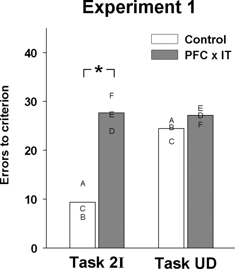
The frontal cortex and inferior temporal cortex are strongly functionally interconnected. Previous experiments on prefrontal function in monkeys have shown that a disconnection of prefrontal cortex from inferior temporal cortex impairs a variety of complex visual learning tasks but leaves simple concurrent object-reward association learning intact. We investigated the possibility that temporal components of visual learning tasks determine the sensitivity of those tasks to prefrontal-temporal disconnection by adding specific temporal components to the concurrent object-reward association learning task. Monkeys with crossed unilateral lesions of prefrontal cortex and inferior temporal cortex were impaired compared with unoperated controls at associating two-item sequences of visual objects with reward. The impairment was specific to the learning of visual sequences, because disconnection was without effect on object-reward association learning for an equivalent delayed reward. This result was replicated in monkeys with transection of the uncinate fascicle, thus determining the anatomical specificity of the dissociation. Previous behavioral results suggest that monkeys represent the two-item serial compound stimuli in a configural manner, similar to the way monkeys represent simultaneously presented compound stimuli. The representation of simultaneously presented configural stimuli depends on the perirhinal cortex. The present experiments show that the representation of serially presented compound stimuli depends on the interaction of prefrontal cortex and inferior temporal cortex. We suggest that prefrontal-temporal disconnection impairs a wide variety of learning tasks because in those tasks monkeys lay down similar temporally complex representations.


Figures

Figure 1.
Figure 1.
Behavioral tasks. Each panel shows a single discrimination problem from each behavioral task. In each testing session, monkeys learned by trial and error which stimuli led to reward. In each experiment, monkeys learned sets of problems from the two tasks in a counterbalanced manner. In task 2I (left), monkeys associated two-item sequences of objects with reward. In task UD (right), monkeys associated single items with reward separated by an unfilled delay of 2 s.
Figure 2.
Figure 2.
Histological reconstructions. A, Intended area of the asymmetric ablations in monkeys with disconnection of prefrontal cortex from inferior temporal cortex (group PFC × IT) shown from lateral, ventral, and medial views. The shaded areas indicate the areas of intended removal; the unilateral prefrontal ablation is shown in light gray, and the unilateral inferior temporal ablation is shown in dark gray. Monkeys received a prefrontal ablation in one hemisphere and an inferotemporal ablation in the opposite hemisphere. The hemisphere (left or right) that received each ablation was counterbalanced across monkeys. PS, Principal sulcus; AS, arcuate sulcus; CIN, cingulate sulcus; ROS, rostral sulcus; MOS, medial orbital sulcus; LOS, lateral orbital sulcus; IOS, inferior occipital sulcus; STS, superior temporal sulcus; LS, lateral sulcus; OTS, occipitotemporal sulcus; AMTS, anterior middle temporal sulcus; RS, rhinal sulcus. B, Coronal sections of the actual and reconstructed lesions of monkeys from group PFC × IT at different anterior–posterior levels [estimated from Paxinos et al. (2000)]. The first column shows four actual sections taken from monkey D. The second column shows a reconstruction, colored in red, of the extent of the asymmetric prefrontal and inferotemporal removal in monkey D represented on cresyl violet-stained sections taken from a normal macaque brain. The remaining two columns show similar reconstructions from monkeys E and F. Monkey D had left hemisphere prefrontal ablation and right hemisphere inferior temporal ablation. Monkeys E and F had right hemisphere prefrontal ablation and left hemisphere inferior temporal ablation.
Figure 3.
Figure 3.
Experiment 1. Learning proficiency in each task is shown by the mean number of errors per set of visual discrimination problems; group means are shown by bars, and individual means are shown by letters. Letters correspond to each monkey's histologically verified lesions shown in Figure 2. Monkeys with disconnection of prefrontal cortex from inferior temporal cortex (shaded bars; PFC × IT) were impaired relative to controls (unshaded bars) at associating two-item sequences of visual objects with reward (Task 2I). The same monkeys were not impaired at associating single items with reward after an unfilled delay (Task UD). *p < 0.05.
Figure 4.
Figure 4.
Experiment 2. a, Coronal sections of the actual and reconstructed lesions in monkeys A–C, who had disconnection of prefrontal and inferotemporal cortex by surgical section of the uncinate fascicle. b, Learning proficiency on each task is shown by the mean number of errors per set of visual discrimination problems; group means are shown by bars, and individual means are shown by letters. Letters correspond to each monkey's histologically verified lesions shown in a. Similar to the monkeys in experiment 1, these monkeys were impaired at associating two-item sequences of visual objects with reward (b, task 2I) but were not impaired at associating single items with reward after an unfilled delay (b, task UD), compared with their preoperative performance (unshaded bars). *p < 0.05.

Similar articles

Cited by

References

    1. Browning PG, Gaffan D. Impairment in object-in-place scene learning after uncinate fascicle section in macaque monkeys. Behav Neurosci. 2008;122:477–482. - PubMed
    1. Browning PGF, Easton A, Buckley MJ, Gaffan D. The role of prefrontal cortex in object-in-place learning in monkeys. Eur J Neurosci. 2005;22:3281–3291. - PubMed
    1. Browning PGF, Easton A, Gaffan D. Frontal-temporal disconnection abolishes object discrimination learning set in macaque monkeys. Cereb Cortex. 2007;17:859–864. - PubMed
    1. Buckley MJ, Gaffan D. Perirhinal cortex ablation impairs configural learning and paired-associate learning equally. Neuropsychologia. 1998;36:535–546. - PubMed
    1. Buckley MJ, Gaffan D, Murray EA. Functional double dissociation between two inferior temporal cortical areas: perirhinal cortex versus middle temporal gyrus. J Neurophysiol. 1997;77:587–598. - PubMed

Publication types

LinkOut - more resources