. 2008 Apr 9;28(15):3976-87.
doi: 10.1523/JNEUROSCI.5692-07.2008.

Prominent role of the spinal central pattern generator in the recovery of locomotion after partial spinal cord injuries

Affiliations

Prominent role of the spinal central pattern generator in the recovery of locomotion after partial spinal cord injuries

Grégory Barrière et al. J Neurosci. .

Abstract

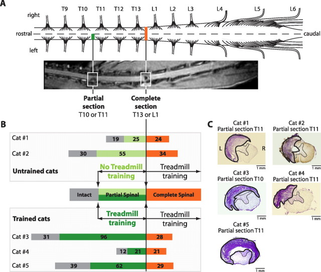
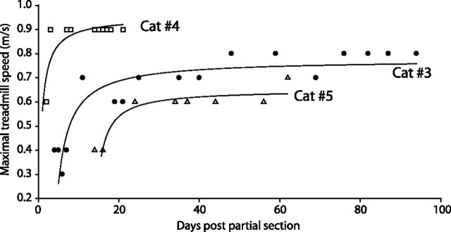
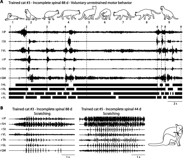
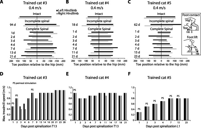
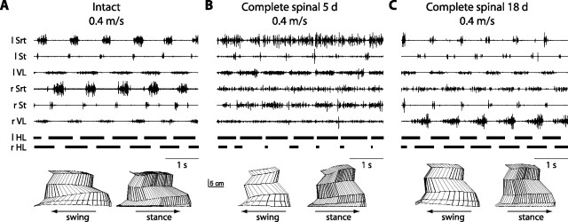
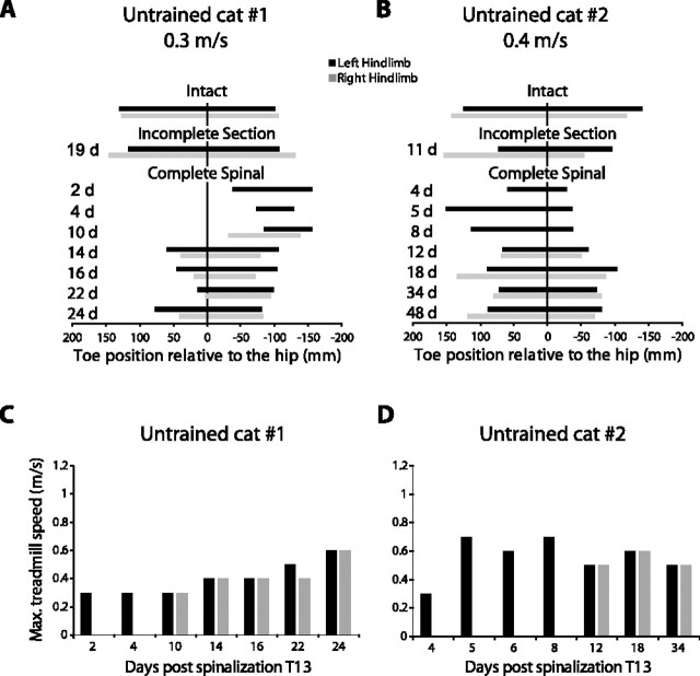
The re-expression of hindlimb locomotion after complete spinal cord injuries (SCIs) is caused by the presence of a spinal central pattern generator (CPG) for locomotion. After partial SCI, however, the role of this spinal CPG in the recovery of hindlimb locomotion in the cat remains mostly unknown. In the present work, we devised a dual-lesion paradigm to determine its possible contribution after partial SCI. After a partial section of the left thoracic segment T10 or T11, cats gradually recovered voluntary quadrupedal locomotion. Then, a complete transection was performed two to three segments more caudally (T13-L1) several weeks after the first partial lesion. Cats that received intensive treadmill training after the partial lesion expressed bilateral hindlimb locomotion within hours of the complete lesion. Untrained cats however showed asymmetrical hindlimb locomotion with the limb on the side of the partial lesion walking well before the other hindlimb. Thus, the complete spinalization revealed that the spinal CPG underwent plastic changes after the partial lesions, which were shaped by locomotor training. Over time, with further treadmill training, the asymmetry disappeared and a bilateral locomotion was reinstated. Therefore, although remnant intact descending pathways must contribute to voluntary goal-oriented locomotion after partial SCI, the recovery and re-expression of the hindlimb locomotor pattern mostly results from intrinsic changes below the lesion in the CPG and afferent inputs.


Figures

Figure 1.
Figure 1.
General description of the dual-lesion paradigm. A, Schematic drawing of a cat spinal cord and corresponding anatomical MRI view (coronal) extracted from cat 5 showing the levels of the incomplete (T10–T11) and complete (T13–L1) sections. B, Sequence of events used in five cats aligned on the time of complete section. Cats were first documented in the intact state (gray) for several weeks after a partial thoracic section on the left side (green) and finally after a complete section (orange) of the spinal cord. Three cats (3, 4, and 5) were intensively trained (dark green) on a treadmill after the partial spinal section for several weeks, whereas cats 1 and 2 were only evaluated (light green) early after the partial section and were not systematically trained. The number of days for each state is indicated in the corresponding horizontal bars for each cat. C, Histology of the partial lesion of the left thoracic segment at T10–T11 in untrained (1 and 2) and trained (3, 4, and 5) cats. The lesioned area is delimited by a continuous black line and the intact gray matter by a dotted line, the remainder corresponding to the intact white matter.
Figure 2.
Figure 2.
Left limb kinematics and bilateral EMG activity of a trained cat (5) walking on the treadmill in the intact state and at different epochs after the incomplete spinal cord lesion. A–D, EMG bursts of selected muscles in the left (l) and right (r) hindlimbs (top). The four bottom traces show associated duty cycles of the stance phases for the HLs and FLs bilaterally. The bottom panels show a stick representation of a typical swing and stance phase of the left hindlimb reconstructed from video recordings.
Figure 3.
Figure 3.
Locomotor performance of trained cats after incomplete spinal section. The maximal speed attained on the treadmill for each trained cat was assessed as a function of days after the partial section.
Figure 4.
Figure 4.
Treadmill training promotes locomotor recovery after incomplete spinal lesion. A, B, Stick figures representing consecutive step cycles of the left hindlimb in the intact state (0.4 m/s) and 11 d after the incomplete spinal lesion in one untrained (2) and one trained (3) cat. The bottom panel shows the corresponding angular excursions of the hip, knee, ankle, and MTP joints 11 d after the partial section. Note that the untrained cat walked at 0.2 m/s 11 d after the partial lesion, whereas the trained cat stepped at 0.4 m/s.
Figure 5.
Figure 5.
Voluntary motor behavior and scratching in trained cats after the incomplete spinal lesion. A, Spontaneous voluntary motor behavior (top) and corresponding EMG activity in cat 3. Vertical doted lines in the electromyogram indicate the time for each figurine. The four bottom traces show the footfall pattern for each limb. B, EMG recordings during scratching of the right leg in cats 3 and 5 at 88 and 44 d post partial section, respectively. The cat figurine on the far right illustrating the scratching was reconstructed from video recordings of cat 5.
Figure 6.
Figure 6.
Left limb kinematics and bilateral EMG activity of a trained cat (4) walking at 0.8 m/s in the intact, incomplete, and complete spinal states. A–C, EMG bursts of selected muscles in the left (l) and right (r) hindlimbs (top). The two bottom traces show associated duty cycles of the stance phases for both HLs. The bottom panels show a stick representation of a typical swing and stance phase of the left hindlimb reconstructed from video recordings.
Figure 7.
Figure 7.
Time course of the recovery of hindlimb locomotion in trained cats after complete spinalization. A–C, Position of the toes relative to the hip for both hindlimbs at foot contact and lift (see insert on the right) at different epochs during the study in trained cats 3 (A), 4 (B), and 5 (C). The left and right extremity for each bar represent the mean position of the toes at foot contact and foot lift, respectively. D–F, The maximal speed attained by each limb for each trained cat as a function of time after the complete spinal section. PS, Perineal stimulation.
Figure 8.
Figure 8.
Left hindlimb kinematics and bilateral EMG activity of an untrained cat (2) walking at 0.4 m/s in the intact state, at 5 and 18 d after the complete spinalization. A–C, EMG bursts of selected muscles in the left (l) and right (r) hindlimbs (top). The two bottom traces show associated duty cycles of the stance phases for both HLs. The bottom panels give a stick representation of typical swing and stance phases of the left hindlimb reconstructed from video recordings.
Figure 9.
Figure 9.
Recovery of hindlimb locomotion after the complete lesion in untrained cats. A, B, Diagrams illustrating the toe position relative to the hip of both hindlimbs at contact and lift at different epochs of the complete dual-lesion paradigm, respectively, in untrained cats 1 (A) and 2 (B). C, D, The maximal speed attained by each limb (performance) for each untrained cat as a function of time after the complete spinal section.

Similar articles

Cited by

References

    1. Ballermann M, Fouad K. Spontaneous locomotor recovery in spinal cord injured rats is accompanied by anatomical plasticity of reticulospinal fibers. Eur J Neurosci. 2006;23:1988–1996. - PubMed
    1. Barbeau H, Rossignol S. Recovery of locomotion after chronic spinalization in the adult cat. Brain Res. 1987;412:84–95. - PubMed
    1. Barbeau H, Rossignol S. Enhancement of locomotor recovery following spinal cord injury. Curr Opin Neurol. 1994;7:517–524. - PubMed
    1. Barbeau H, Visintin M. Optimal outcomes obtained with body-weight support combined with treadmill training in stroke subjects. Arch Phys Med Rehabil. 2003;84:1458–1465. - PubMed
    1. Bareyre FM, Kerschensteiner M, Raineteau O, Mettenleiter TC, Weinmann O, Schwab ME. The injured spinal cord spontaneously forms a new intraspinal circuit in adult rats. Nat Neurosci. 2004;7:269–277. - PubMed

Publication types