. 2008 Jun;19(6):2588-96.
doi: 10.1091/mbc.e08-02-0187. Epub 2008 Apr 9.

R-Spondin family members regulate the Wnt pathway by a common mechanism

Affiliations

R-Spondin family members regulate the Wnt pathway by a common mechanism

Kyung-Ah Kim et al. Mol Biol Cell. 2008 Jun.

Abstract

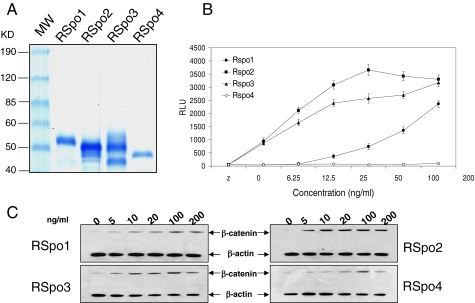
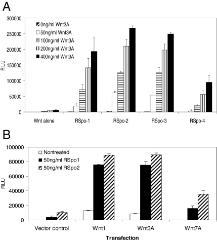
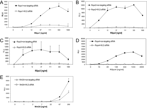
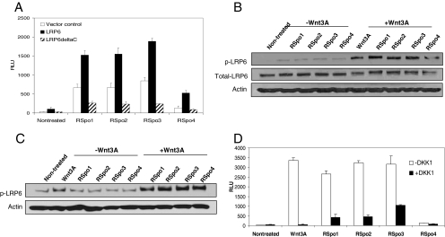
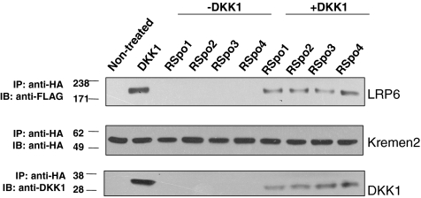
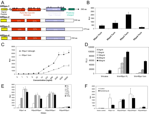
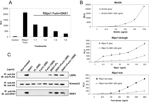
The R-Spondin (RSpo) family of secreted proteins is implicated in the activation of the Wnt signaling pathway. Despite the high structural homology between the four members, expression patterns and phenotypes in knockout mice have demonstrated striking differences. Here we dissected and compared the molecular and cellular function of all RSpo family members. Although all four RSpo proteins activate the canonical Wnt pathway, RSpo2 and 3 are more potent than RSpo1, whereas RSpo4 is relatively inactive. All RSpo members require Wnt ligands and LRP6 for activity and amplify signaling of Wnt3A, Wnt1, and Wnt7A, suggesting that RSpo proteins are general regulators of canonical Wnt signaling. Like RSpo1, RSpo2-4 antagonize DKK1 activity by interfering with DKK1 mediated LRP6 and Kremen association. Analysis of RSpo deletion mutants indicates that the cysteine-rich furin domains are sufficient and essential for the amplification of Wnt signaling and inhibition of DKK1, suggesting that Wnt amplification by RSpo proteins may be a direct consequence of DKK1 inhibition. Together, these findings indicate that RSpo proteins modulate the Wnt pathway by a common mechanism and suggest that coexpression with specific Wnt ligands and DKK1 may determine their biological specificity in vivo.


Figures

Figure 1.
Figure 1.
RSpo family proteins activate β-catenin signaling. (A) Coomassie-stained RSpo 1, 2, 3, and 4 proteins. Purified Rspo proteins were resolved on a 4–20% SDS-PAGE gel under reducing conditions and were visualized by Coomassie blue staining. Each RSpo lane contains 500 ng of the purified protein. The first lane contains markers with molecular masses as indicated at the left. Estimated purities for RSpo1, 2, 3, and 4 are 98, 90, 93, and 85%, respectively, based on immunoreactive bands in Western blotting using antiV5 antibodies. (B) Activities of RSpo proteins were measured by TCF reporter assay. HEK293 cells stably transfected with a 16-TCF-luciferase reporter construct were treated with increasing concentrations of RSpo proteins for 20 h, and reporter activity was measured. Data represent relative light units (RLU). Assays were done in triplicate and the results are mean ± SD. (C) β-Catenin stabilization was measured by Western blotting using an anti-β-catenin antibody. HEK293 cells were treated with various concentrations of RSpo proteins for 3 h, and the cytosolic fractions were prepared for immunoblotting for β-catenin. β-Actin was used as a loading control.
Figure 2.
Figure 2.
RSpo proteins synergize with various Wnt proteins in TCF-mediated transcriptional activation. (A) HEK293 cells stably transfected with a 16-TCF-luciferase reporter construct were treated with increasing concentrations of Wnt3A protein in the absence or presence of 20 ng/ml recombinant RSpo proteins. After a 20-h incubation, reporter activity was measured using a luminometer. (B) Stable 293 TCF reporter cells were transfected with Wnt1, Wnt3A, or Wnt7A expression constructs in 96-well (50 ng per transfection). Twenty four hours after the transfection, cells were treated with either RSpo1 or RSpo2 at 50 ng/ml and TCF reporter activity was measured 20 h after the incubation. Empty vector was used as a transfection control.
Figure 3.
Figure 3.
Activities of RSpo proteins are dependent on an endogenous pool of Wnt ligands. Stable HEK-293 reporter cells were transfected with either nontargeting control siRNA or WLS siRNA duplexes (10 nM), treated with indicated amounts of RSpo1 (A), RSpo2 (B), RSpo3 (C), RSpo4 (D), or Wnt3A (E) proteins. Reporter activity was analyzed as described above.
Figure 4.
Figure 4.
LRP6 is implicated in RSpo-mediated β-catenin signaling. (A) HEK293 cells stably expressing 16-TCF reporter were transfected with either wt LRP6 or LRP6ΔC expression constructs, treated with indicated RSpo proteins (200 ng/ml), and the luciferase reporter activity was determined 20 h after the incubation. Empty vector was used as a transfection control. (B) Regular HEK293 cells were treated with RSpo proteins (200 ng/ml), Wnt3A (200 ng/ml), or Wnt3A (200 ng/ml), and individual RSpo protein (200 ng/ml) together for 4 h. Total cell lysates were prepared and resolved by SDS-PAGE. Phosphorylation of endogenous LRP6 was detected by immunoblotting using anti-phospho-LRP6 antibodies. (C) HEK293 cells were stably transfected with a LRP6-FLAG expression construct and treated with Wnt3A (200 ng/ml), RSpo proteins alone (200 ng/ml) or Wnt3A and RSpo together for 4 h. The total cell lysates were prepared and resolved by SDS-PAGE followed by immunoblotting using anti-phospho-LRP6 antibodies. (D) Stable HEK293 A6 cells were treated with Wnt3A (250 ng/ml) or RSpo protein alone (125 ng/ml) or in combination with recombinant DKK1 protein (400 ng/ml). The luciferase reporter activity was determined 20 h after incubation.
Figure 5.
Figure 5.
RSpo proteins antagonize DKK1-mediated interaction of Kremen2 and LRP6. HEK-293 cells, stably expressing LRP6-FLAG, were transfected with Kremen2-HA and treated with DKK-1 (100 ng/ml) in the presence or absence of indicated RSpo proteins (200 ng/ml) for 30 min. The total cell lysates were immunoprecipitated with anti-HA antibody for Kremen2 and coimmunoprecipitated LRP6, or DKK1 proteins were immunoblotted with anti-FLAG or anti-DKK1 antibodies, respectively. DKK1 induced an interaction of Kremen2 and LRP6 proteins and RSpo proteins reduced the DKK1-mediated Kremen2 and LRP6 interaction.
Figure 6.
Figure 6.
Furin domains confer the relative activity of RSpo proteins. (A) Domain diagrams of RSpo family members. (B) The furin domain of each RSpo family member was cloned and transfected into HEK293 cells stably transfected with TCF reporter construct. Activation of luciferase reporter was determined by a luminometer. (C) Activities of purified full-length and the furin domain of RSpo1 protein were tested by TCF reporter assay. Stable HEK293 TCF reporter cells were treated with increasing concentrations of each protein for 20 h, and TCF transcriptional activity was measured. (D) The furin domain amplifies Wnt3A activity. Stable HEK293 cells were treated with varying concentrations of Wnt3A in the presence of 20 ng/ml RSpo1 full-length protein or furin domain. (E) To examine whether furin domains are responsible for the relative activities of RSpo proteins, various chimeras were generated, in which the furin domain of RSpo1 gene was replaced with those of RSpo2, 3, or 4. Each chimeric construct was transfected into stable HEK293 TCF reporter cells with indicated amounts of DNA in 96-well plates, and TCF reporter activity was measured 48 h after transfection. (F) Specific activities of RSpo chimeras were tested using an RSpo1-specific neutralizing mAb. Cells were treated with 1 μg/ml neutralizing antibody after transfection of stable reporter cells with each chimeric construct, and reporter activity was measured.
Figure 7.
Figure 7.
The furin domain mediates RSpo-dependent inhibition of DKK1 activity. (A) Stable 293 TCF reporter cells were treated with purified RSpo1 furin domain (100 ng/ml) alone or in combination with increasing concentrations of DKK1 protein (ratios of amounts of RSpo furin domain: DKK1 are shown). TCF reporter activity was measured 20 h after the incubation. (B) Stable 293 TCF reporter cells were treated with DKK1 protein (400 ng/ml) in the presence of increasing amounts of Wnt3A (top panel), RSpo1 full-length (middle panel) or RSpo furin domain (bottom panel) proteins, and TCF reporter activity was determined. (C) To examine the effect of the furin domain on LRP6 and Kremen interaction, coimmunoprecipitation was performed. HEK293 cells stably transfected with a FLAG-tagged LRP6 expression construct, were transfected with HA-tagged hKremen2. Cells were treated as indicated, and total cell lysates were used to immunoprecipitate Kremen2 using anti-HA antibodies. Coimmunoprecipitated LRP6 or DKK1 were detected using anti-FLAG or anti-DKK1 antibodies, respectively. The representative blots are shown.

References

    1. Aoki M., Mieda M., Ikeda T., Hamada Y., Nakamura H., Okamoto H. R-spondin3 is required for mouse placental development. Dev. Biol. 2006;301:218–226. - PubMed
    1. Banziger C., Soldini D., Schutt C., Zipperlen P., Hausmann G., Basler K. Wntless, a conserved membrane protein dedicated to the secretion of Wnt proteins from signaling cells. Cell. 2006;125:509–522. - PubMed
    1. Behrens J., Jerchow B. A., Wurtele M., Grimm J., Asbrand C., Wirtz R., Kuhl M., Wedlich D., Birchmeier W. Functional interaction of an axin homolog, conductin, with beta-catenin, APC, and GSK3beta. Science. 1998;280(5363):596–599. - PubMed
    1. Bejsovec A. Wnt pathway activation: new relations and locations. Cell. 2005;120:11–14. - PubMed
    1. Bell S. M., Schreiner C. M., Hess K. A., Anderson K. P., Scott W. J. Asymmetric limb malformations in a new transgene insertional mutant, footless. Mech. Dev. 2003;120:597–605. - PubMed

LinkOut - more resources