. 2008 May 1;94(9):3363-83.
doi: 10.1529/biophysj.107.114058.

Diffusion of the second messengers in the cytoplasm acts as a variability suppressor of the single photon response in vertebrate phototransduction

Affiliations

Diffusion of the second messengers in the cytoplasm acts as a variability suppressor of the single photon response in vertebrate phototransduction

Paolo Bisegna et al. Biophys J. .

Abstract

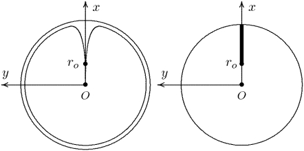
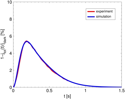
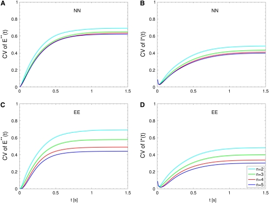
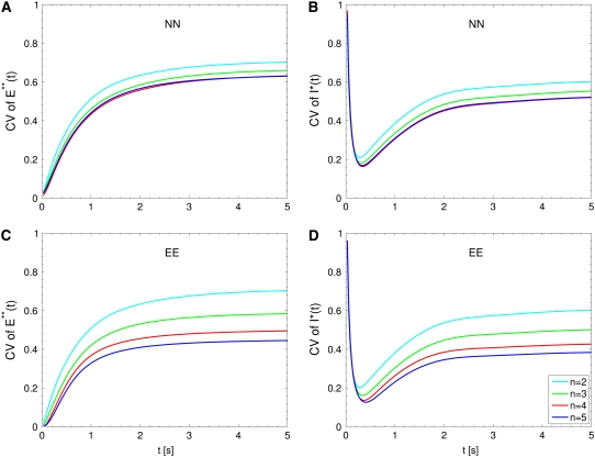
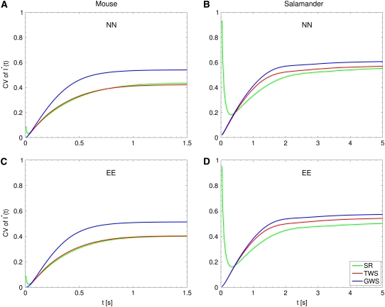
The single photon response in vertebrate phototransduction is highly reproducible despite a number of random components of the activation cascade, including the random activation site, the random walk of an activated receptor, and its quenching in a random number of steps. Here we use a previously generated and tested spatiotemporal mathematical and computational model to identify possible mechanisms of variability reduction. The model permits one to separate the process into modules, and to analyze their impact separately. We show that the activation cascade is responsible for generation of variability, whereas diffusion of the second messengers is responsible for its suppression. Randomness of the activation site contributes at early times to the coefficient of variation of the photoresponse, whereas the Brownian path of a photoisomerized rhodopsin (Rh*) has a negligible effect. The major driver of variability is the turnoff mechanism of Rh*, which occurs essentially within the first 2-4 phosphorylated states of Rh*. Theoretically increasing the number of steps to quenching does not significantly decrease the corresponding coefficient of variation of the effector, in agreement with the biochemical limitations on the phosphorylated states of the receptor. Diffusion of the second messengers in the cytosol acts as a suppressor of the variability generated by the activation cascade. Calcium feedback has a negligible regulatory effect on the photocurrent variability. A comparative variability analysis has been conducted for the phototransduction in mouse and salamander, including a study of the effects of their anatomical differences such as incisures and photoreceptors geometry on variability generation and suppression.


Figures

FIGURE 1
FIGURE 1
(Left) Transversal cross section of the ROS bearing an incisure. (Right) Limit of such a cross section, bearing the limiting incisure V.
FIGURE 2
FIGURE 2
The red curve reports the average of an extensive set of experimental SPR responses for mouse, kindly provided to us by F. Rieke. The blue curve is our numerical simulation of the mouse SPR using the mathematical model of Appendix A for the set of parameters in Table 3. The agreement is excellent, thereby showing that this selection parameters accurately reflects the timecourse and amplitude of experimentally generated light responses.
FIGURE 3
FIGURE 3
Mouse: comparing the CV of the total activated effectors formula image at time t with the CV of the total relative charge formula image up to time t. (NN) Shutoff of Rh* in n biochemical states of decreasing duration and catalytic activity (see Biochemical Sequences {τj} and {νj}). (EE) Shutoff of Rh* in n theoretical states of equal duration and equal catalytic activity (see Sequence for Which τjνj = Const). All simulations assume all activation steps as random (Case 4 of Random Events Contributing to SPR Variability). In all cases, CV decreases with increasing n. (A and B) For the biochemical state NN, the CV of both E**(t) and I*(t) stabilizes asymptotically after 3–4 phosphorylated states. A CV of ∼60% for E**(t) at times past the peak time is reduced to a CV of ∼40% for the corresponding photocurrent I*(t). (C and D) For the theoretical state EE, increasing n gives in all cases a decreased CV although at a decreasing rate for increasing n. The CV comparison E**(t) (∼60%) to I*(t) (∼40%) is still present, thus pointing to an intrinsic variability reduction effect of the diffusion part of the process.
FIGURE 4
FIGURE 4
Salamander: comparing the CV of the total activated effectors formula image at time t with the CV of the total relative charge formula image up to time t. (NN) Shutoff of Rh* in n biochemical states of decreasing duration and catalytic activity (see Biochemical Sequences {τj} and {νj}); (EE) Shutoff of Rh* in n theoretical states of equal duration and equal catalytic activity (see Sequence for Which τjνj = Const). All simulations assume all activation steps as random (Case 4 of Random Events Contributing to SPR Variability). In all cases, CV decreases with increasing n. For NN, the CV stabilizes for n ≥ 3 and it is essentially the same for n = 3, 4, 5. For EE, the CV keeps decreasing with increasing n, although at a decreasing rate for increasing n. For the salamander at early times, the CV is initially large and then rapidly drops. No similar effect occurs in mouse. (A and B) For the biochemical state NN, the CV of both E**(t) and I*(t) stabilizes asymptotically after 3–4 phosphorylated states. A CV of ∼60% for E**(t) at times past the peak time is reduced to a CV of ∼50% for the corresponding photocurrent I*(t). (C and D) For the theoretical state EE, increasing n gives in all cases a decreased CV although at a decreasing rate for increasing n. The CV comparison E**(t) (∼60%) to I*(t) (∼50%) is still present, thus pointing to an intrinsic variability reduction effect of the diffusion part of the process. The suppression of CV for the photocurrent I*(t) with respect to CV of the activating E**(t), while present, is less dramatic than for mouse (Fig. 3, A and B). In addition, we observe a sharp variability at early times, which is likely due to presence of the incisures and their distributed geometry. This is supported by the absence of such incipient CV, in lumped models insensitive to incisures geometry (see also captions of Fig. 5). This early-time high CV seems also to be due to the random position of the activation site. Indeed, photons absorbed close to the disk boundary, yield a faster response than those absorbed far away from the boundary, say near the center of the disk. After a short time, depending on the diffusivity coefficients on the disk and on the disk radius, this difference is reduced. In the mouse, where diffusivities are larger and the radius is smaller than similar parameters in the salamander, no significant increase of variability at early times is observed (see also Fig. 3).
FIGURE 5
FIGURE 5
CV of formula image: total relative charge up to time t. (SR) Space-resolved; (TWS) transversally well-stirred; (GWS) globally well-stirred; (NN) shutoff of Rh* in n biochemical states of decreasing duration and catalytic activity (see Biochemical Sequences {τj} and {νj}); and (EE) shutoff of Rh* in n theoretical states of equal duration and equal catalytic activity (see Sequence for Which τjνj = Const). All simulations assume all activation steps as random (Case 4 of Random Events Contributing to SPR Variability). In all cases, the CV computed with the GWS model is higher than the one computed with the other two models. A high CV at early times for the salamander, detected by the SR model (where geometry and incisures matter), is not detected by the lumped models TWS and GWS. This suggests the interpretation that a high CV at the inception of the activation cascade is due to the architecture of the salamander ROS.

References

    1. Aho, A. C., K. Donner, C. Hyden, L. O. Larsen, and T. Reuter. 1988. Low retinal noise in animals with low body temperature allows high visual sensitivity. Nature. 334:348–350. - PubMed
    1. Hecht, S., S. Shlaer, and M. Pierenne. 1942. Energy, quanta and vision. J. Gen. Physiol. 25:819–840. - PMC - PubMed
    1. Baylor, D. A., T. D. Lamb, and K. W. Yau. 1979. The membrane current of single rod outer segments. J. Physiol. 288:589–611. - PMC - PubMed
    1. Baylor, D. A., T. D. Lamb, and K. W. Yau. 1979. Responses of retinal rods to single photons. J. Physiol. 288:613–634. - PMC - PubMed
    1. Felber, S., H. P. Breuer, F. Petruccione, J. Honerkamp, and K. P. Hofmann. 1996. Stochastic simulation of the transducin GTPase cycle. Biophys. J. 71:3051–3063. - PMC - PubMed

Publication types

LinkOut - more resources