

Abnormal TDP-43 immunoreactivity in AD modifies clinicopathologic and radiologic phenotype
- PMID: 18401022
- PMCID: PMC2779031
- DOI: 10.1212/01.wnl.0000304041.09418.b1
Abnormal TDP-43 immunoreactivity in AD modifies clinicopathologic and radiologic phenotype
Abstract
Background: TAR DNA-binding protein 43 (TDP-43) is one of the major disease proteins in frontotemporal lobar degeneration with ubiquitin immunoreactivity. Approximately one-fourth of subjects with pathologically confirmed Alzheimer disease (AD) have abnormal TDP-43 (abTDP-43) immunoreactivity. The aim of this study was to determine whether subjects with pathologically confirmed AD and abTDP-43 immunoreactivity have distinct clinical, neuropsychological, imaging, or pathologic features compared with subjects with AD without abTDP-43 immunoreactivity.
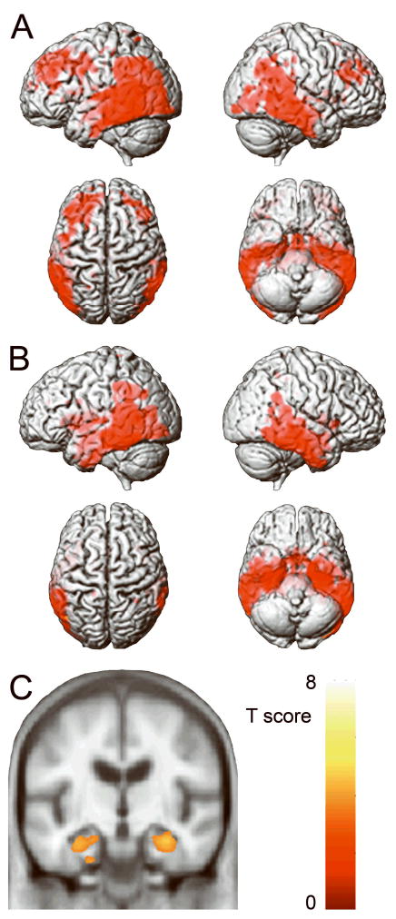
Methods: Eighty-four subjects were identified who had a pathologic diagnosis of AD, neuropsychometric testing, and volumetric MRI. Immunohistochemistry for TDP-43 was performed on sections of hippocampus and medial temporal lobe, and positive cases were classified into one of three types. Neuropsychometric data were collated and compared in subjects with and without abTDP-43 immunoreactivity. Voxel-based morphometry was used to assess patterns of gray matter atrophy in subjects with and without abTDP-43 immunoreactivity compared with age- and sex-matched controls.
Results: Twenty-nine (34%) of the 84 AD subjects had abTDP-43 immunoreactivity. Those with abTDP-43 immunoreactivity were older at onset and death and performed worse on the Clinical Dementia Rating scale, Mini-Mental State Examination, and Boston Naming Test than subjects without abTDP-43 immunoreactivity. Subjects with and without abTDP-43 immunoreactivity had medial temporal and temporoparietal gray matter loss compared with controls; however, those with abTDP-43 immunoreactivity showed greater hippocampal atrophy. Multivariate logistic regression adjusting for age at death demonstrated that hippocampal sclerosis was the only pathologic predictor of abTDP-43 immunoreactivity.
Conclusions: The presence of abnormal TDP-43 immunoreactivity is associated with a modified Alzheimer disease clinicopathologic and radiologic phenotype.
Figures

References
-
- Josephs KA, Holton JL, Rossor MN, et al. Frontotemporal lobar degeneration and ubiquitin immunohistochemistry. Neuropathol Appl Neurobiol. 2004;30:369–373. - PubMed
-
- Neumann M, Sampathu DM, Kwong LK, et al. Ubiquitinated TDP-43 in frontotemporal lobar degeneration and amyotrophic lateral sclerosis. Science. 2006;314:130–133. - PubMed
-
- Josephs KA, Petersen RC, Knopman DS, et al. Clinicopathologic analysis of frontotemporal and corticobasal degenerations and PSP. Neurology. 2006;66:41–48. - PubMed
-
- Whitwell JL, Josephs KA, Rossor MN, et al. Magnetic resonance imaging signatures of tissue pathology in frontotemporal dementia. Arch Neurol. 2005;62:1402–1408. - PubMed
Publication types
MeSH terms
Substances
Grants and funding
LinkOut - more resources
Full Text Sources
Other Literature Sources
Medical