. 2008 Apr 10;452(7188):759-63.
doi: 10.1038/nature06859.

Endothelins are vascular-derived axonal guidance cues for developing sympathetic neurons

Affiliations

Endothelins are vascular-derived axonal guidance cues for developing sympathetic neurons

Takako Makita et al. Nature. .

Abstract

During development, sympathetic neurons extend axons along a myriad of distinct trajectories, often consisting of arteries, to innervate one of a large variety of distinct final target tissues. Whether or not subsets of neurons within complex sympathetic ganglia are predetermined to innervate select end-organs is unknown. Here we demonstrate in mouse embryos that the endothelin family member Edn3 (ref. 1), acting through the endothelin receptor EdnrA (refs 2, 3), directs extension of axons of a subset of sympathetic neurons from the superior cervical ganglion to a preferred intermediate target, the external carotid artery, which serves as the gateway to select targets, including the salivary glands. These findings establish a previously unknown mechanism of axonal pathfinding involving vascular-derived endothelins, and have broad implications for endothelins as general mediators of axonal growth and guidance in the developing nervous system. Moreover, they suggest a model in which newborn sympathetic neurons distinguish and choose between distinct vascular trajectories to innervate their appropriate end organs.


Figures
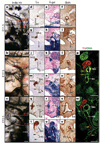
Figure 1
Figure 1. Development of vascular and sympathetic systems
a–c, India ink visualization of the carotid arteries. The red lines indicate the approximate levels of sections shown in d–w. d–w, Serial transverse sections at the approximate levels indicated in panels a–c were stained with X-gal or immunostained for TH or alpha smooth muscle actin. d–f, j–l and q–s are sections through the internal carotid artery. g–i,m–p and t–w are at the level of the carotid bifurcation and show the base of the internal and external carotid arteries.m, t, Arrows denote SCG axons extending along the external carotid artery. Low-magnification views of the same sections are provided in Supplementary Fig. 1. At least ten embryos at each stage were analysed in this manner with comparable results. p, w, Confocal immunofluorescent images of sections of E12.5 and E13.5 embryos at a level approximately as indicated in b and c, respectively, showing TH-positive axons (red) projecting along the smooth muscle layer (green) of the base of internal and external carotid arteries (yellow arrows). Abbreviations: 3, 4 and 6, third, fourth and sixth pharyngeal arch arteries, respectively; bic, base of internal carotid artery; cc, common carotid artery; dAo, dorsal aorta; ec, external carotid artery; ic, internal carotid artery; pec, primitive external carotid artery; pic, primitive internal carotid artery; sa, stapedial artery; sc, sympathetic chain; and scg, superior cervical ganglion. Scale bars, 100 µm.
Figure 2
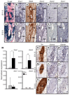
Figure 2. Expression of endothelin signalling components
a–l, Serial frontal sections of E11.5 (a–f) and E12.5 (g–l) Wnt1Cre/R26R embryos, stained with X-gal, immunostained for SMA, or digoxygenin-labelled by in situ hybridization for the indicated genes. m, Quantitative real-time PCR analysis. Total RNA was extracted from dissected vascular segments from five individual E12.5 wild-type embryos isolated from two different litters. Total RNA was reverse transcribed, and was evaluated for gene expression by quantitative real-time PCR as described in the Methods. Expression levels were normalized to the amount of GAPDH transcripts and are scaled as absolute values. ic and ec, internal and external carotid artery segments. The grey bars represent the signal associated with the internal carotid arteries, whereas the black bars represent the signal associated with the external carotid arteries. ND, not detected. Error bars, mean ± s.e.m.; *P<0.01; **P<0.005. n–v, Serial frontal sections of embryos at the indicated stages immunolabelled for TH or labelled by in situ hybridization for Ednra and Ednrb. Sections shown at E11.5 are through the rostral end of the sympathetic chain, and at E12.5 and E13.5 are through the SCG. Results were confirmed with sections from at least five embryos of each stage. Scale bars, 100 µm.
Figure 3
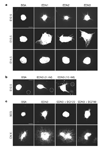
Figure 3. Endothelins promote SCG neurite outgrowth in vitro
a, SCGs from embryos at the indicated stages were harvested and grown in collagen gels prepared with the indicated endothelins (10 nM) or BSA. The average amount of neurite outgrowth from seven separate experiments is shown in Supplementary Fig. 3a. b, Heparin–agarose beads soaked in the indicated concentration of EDN3 or BSA were placed adjacent to E12.5 SCG. Dotted circles indicate the location of the beads. The extent of directional outgrowth was assessed by the relative amount of outgrowth in the proximal direction relative to the distal direction of the beads, as shown in Supplementary Fig. 3b. c, E12.5 SCG (top row) or vagal (bottom row) explants were grown in collagen gels in the presence of EDN3 (5 nM) and the EdnrA antagonist BQ123 (1 µM) or the EdnrB antagonist BQ788 (100 nM) as indicated. Quantification of EDN3-induced neurite outgrowth influenced by each antagonist is shown in Supplementary Fig. 3c, d. Scale bars, 100 µm.
Figure 4
Figure 4. Edn3 and its receptor EdnrA are essential for SCG axonal projections along the external but not internal carotid arteries in mouse embryos
a–l, Histological sections stained with TH or SMA from an E13.5 Ednra−/− embryo and a littermate control. Three mutant embryos analysed from multiple litters exhibited a complete absence of axons along the external carotid artery, similar to the embryo shown in g–l.m–r, Histological sections from an E13.5 Edn3−/− embryo and a littermate control stained with TH. Seven Edn3−/− embryos analysed at E13.5 or older from multiple litters had defects in axonal projections towards the external carotid arteries. Sections shown in the top row are all at the cochlea–pituitary level, and show the rostral projection from the SCG along the internal carotid artery. Shown in the middle row are sections at the level of the branch of the stapedial artery from the internal carotid artery; these reveal the cranial-most extent of the SCG and the initial projection from the SCG to the internal carotid artery. Sections in the lower row were taken at a level immediately above the bifurcation of the external and internal carotid arteries from the common carotid artery. Arrows point to the presence (in panels c and o) or the absence (in i and r) of axonal projections to the external carotid artery. Abbreviations: co, cochlea; pt, pituitary gland; V, trigeminal ganglion; *, histological artefact. Scale bar, 100 µm.

References

    1. Inoue A, et al. The human endothelin family: three structurally and pharmacologically distinct isopeptides predicted by three separate genes. Proc. Natl Acad. Sci. USA. 1989;86:2863–2867. - PMC - PubMed
    1. Arai H, Hori S, Aramori I, Ohkubo H, Nakanishi S. Cloning and expression of a cDNA encoding an endothelin receptor. Nature. 1990;348:730–732. - PubMed
    1. Sakurai T, et al. Cloning of a cDNA encoding a non-isopeptide-selective subtype of the endothelin receptor. Nature. 1990;348:732–735. - PubMed
    1. Schotzinger RJ, Landis SC. Cholinergic phenotype developed by noradrenergic sympathetic neurons after innervation of a novel cholinergic target in vivo. Nature. 1988;335:637–639. - PubMed
    1. Hiruma T, Nakajima Y, Nakamura H. Development of pharyngeal arch arteries in early mouse embryo. J. Anat. 2002;201:15–29. - PMC - PubMed

Publication types

Associated data