

Overexpression of Syk tyrosine kinase in peripheral T-cell lymphomas
- PMID: 18401419
- PMCID: PMC2778211
- DOI: 10.1038/leu.2008.77
Overexpression of Syk tyrosine kinase in peripheral T-cell lymphomas
Abstract
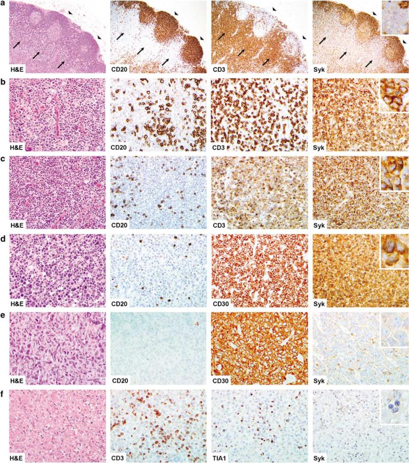
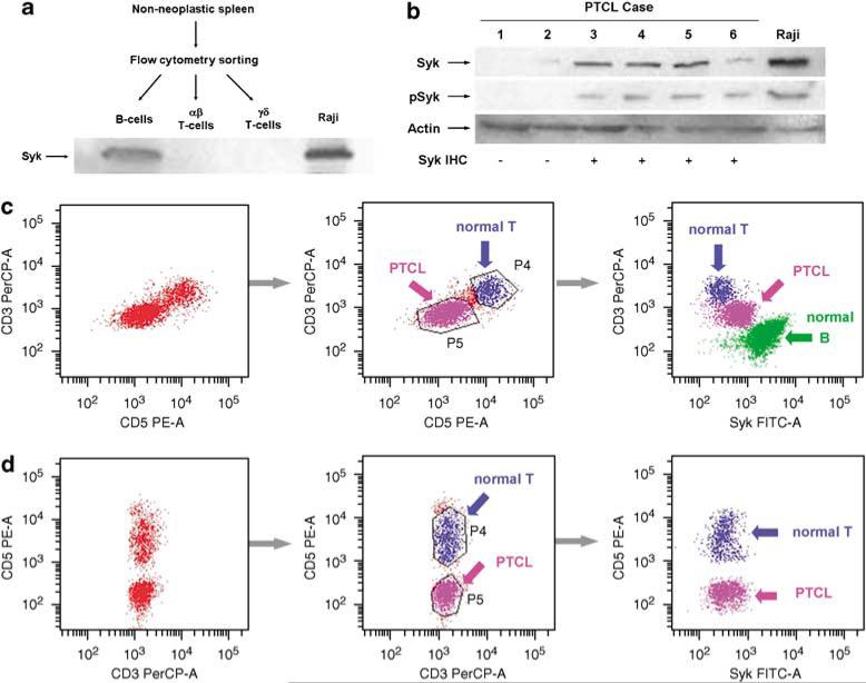
Peripheral T-cell lymphomas (PTCLs) are fatal in the majority of patients and novel treatments, such as protein tyrosine kinase (PTK) inhibition, are needed. The recent finding of SYK/ITK translocations in rare PTCLs led us to examine the expression of Syk PTK in 141 PTCLs. Syk was positive by immunohistochemistry (IHC) in 133 PTCLs (94%), whereas normal T cells were negative. Western blot on frozen tissue (n=6) and flow cytometry on cell suspensions (n=4) correlated with IHC results in paraffin. Additionally, western blot demonstrated that Syk-positive PTCLs show tyrosine (525/526) phosphorylation, known to be required for Syk activation. Fluorescence in situ hybridization showed no SYK/ITK translocation in 86 cases. Overexpression of Syk, phosphorylation of its Y525/526 residues and the availability of orally available Syk inhibitors suggest that Syk merits further evaluation as a candidate target for pharmacologic PTK inhibition in patients with PTCL.
Figures
Similar articles
-
Novel t(5;9)(q33;q22) fuses ITK to SYK in unspecified peripheral T-cell lymphoma.Leukemia. 2006 Feb;20(2):313-8. doi: 10.1038/sj.leu.2404045. Leukemia. 2006. PMID: 16341044
-
The fusion kinase ITK-SYK mimics a T cell receptor signal and drives oncogenesis in conditional mouse models of peripheral T cell lymphoma.J Exp Med. 2010 May 10;207(5):1031-44. doi: 10.1084/jem.20092042. Epub 2010 May 3. J Exp Med. 2010. PMID: 20439541 Free PMC article.
-
Phosphatidylinositol-3-kinase-dependent phosphorylation of SLP-76 by the lymphoma-associated ITK-SYK fusion-protein.Biochem Biophys Res Commun. 2009 Dec 18;390(3):892-6. doi: 10.1016/j.bbrc.2009.10.070. Epub 2009 Oct 20. Biochem Biophys Res Commun. 2009. PMID: 19850008
-
Anaplastic large cell lymphoma: one or more entities among T-cell lymphoma?Hematol Oncol. 2009 Dec;27(4):161-70. doi: 10.1002/hon.897. Hematol Oncol. 2009. PMID: 19358142 Review.
-
The t(2;5) in human lymphomas.Leuk Lymphoma. 1998 Apr;29(3-4):249-56. doi: 10.3109/10428199809068562. Leuk Lymphoma. 1998. PMID: 9684923 Review.
Cited by
-
Enforced expression of Lin28b leads to impaired T-cell development, release of inflammatory cytokines, and peripheral T-cell lymphoma.Blood. 2012 Aug 2;120(5):1048-59. doi: 10.1182/blood-2012-01-401760. Epub 2012 Jun 21. Blood. 2012. PMID: 22723554 Free PMC article.
-
Getting Syk: spleen tyrosine kinase as a therapeutic target.Trends Pharmacol Sci. 2014 Aug;35(8):414-22. doi: 10.1016/j.tips.2014.05.007. Epub 2014 Jun 26. Trends Pharmacol Sci. 2014. PMID: 24975478 Free PMC article. Review.
-
Genetic profiling and biomarkers in peripheral T-cell lymphomas: current role in the diagnostic work-up.Mod Pathol. 2022 Mar;35(3):306-318. doi: 10.1038/s41379-021-00937-0. Epub 2021 Sep 28. Mod Pathol. 2022. PMID: 34584212 Review.
-
Genetic alterations in systemic nodal and extranodal non-cutaneous lymphomas derived from mature T cells and natural killer cells.Cancer Sci. 2012 Aug;103(8):1397-404. doi: 10.1111/j.1349-7006.2012.02321.x. Epub 2012 Jun 26. Cancer Sci. 2012. PMID: 22568409 Free PMC article.
-
Peripheral T cell lymphomas: from the bench to the clinic.Nat Rev Cancer. 2020 Jun;20(6):323-342. doi: 10.1038/s41568-020-0247-0. Epub 2020 Apr 6. Nat Rev Cancer. 2020. PMID: 32249838 Review.
References
-
- Savage KJ, Chhanabhai M, Gascoyne RD, Connors JM. Characterization of peripheral T-cell lymphomas in a single North American institution by the WHO classification. Ann Oncol. 2004;15:1467–1475. - PubMed
-
- Streubel B, Vinatzer U, Willheim M, Raderer M, Chott A. Novel t(5;9)(q33;q22) fuses ITK to SYK in unspecified peripheral T-cell lymphoma. Leukemia. 2006;20:313–318. - PubMed
-
- de Leval L, Savilo E, Longtine J, Ferry JA, Harris NL. Peripheral T-cell lymphoma with follicular involvement and a CD4+/bcl-6+ phenotype. Am J Surg Pathol. 2001;25:395–400. - PubMed
-
- Pogue SL, Kurosaki T, Bolen J, Herbst R. B cell antigen receptor-induced activation of Akt promotes B cell survival and is dependent on Syk kinase. J Immunol. 2000;165:1300–1306. - PubMed
-
- Rinaldi A, Kwee I, Taborelli M, Largo C, Uccella S, Martin V, et al. Genomic and expression profiling identifies the B-cell associated tyrosine kinase Syk as a possible therapeutic target in mantle cell lymphoma. Br J Haematol. 2006;132:303–316. - PubMed
Publication types
MeSH terms
Substances
Grants and funding
LinkOut - more resources
Full Text Sources
Other Literature Sources
Miscellaneous
