. 2008 Sep;15(18):1257-70.
doi: 10.1038/gt.2008.58. Epub 2008 Apr 10.

Inflammatory tumour cell killing by oncolytic reovirus for the treatment of melanoma

Affiliations

Inflammatory tumour cell killing by oncolytic reovirus for the treatment of melanoma

F Errington et al. Gene Ther. 2008 Sep.

Abstract

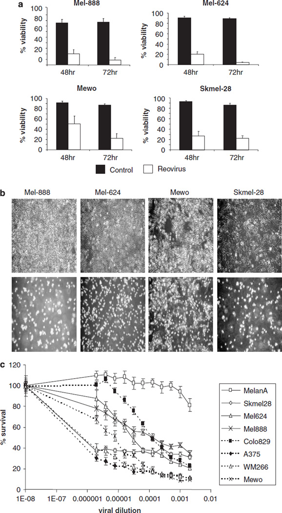
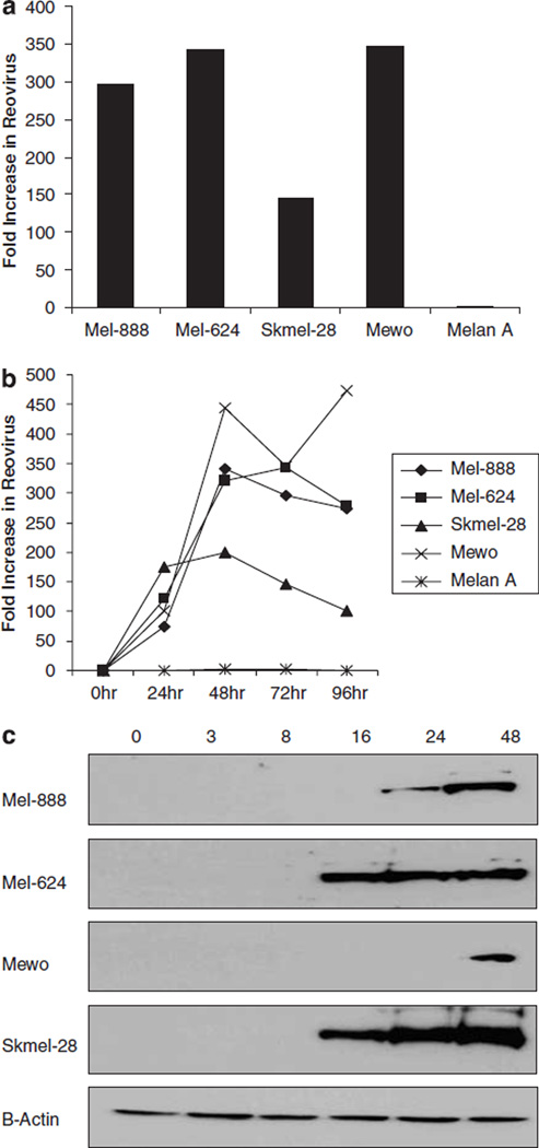
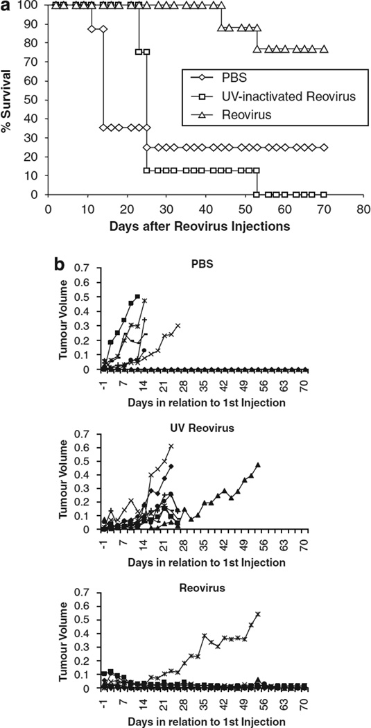
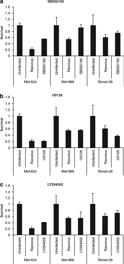
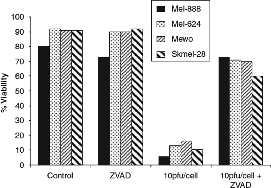
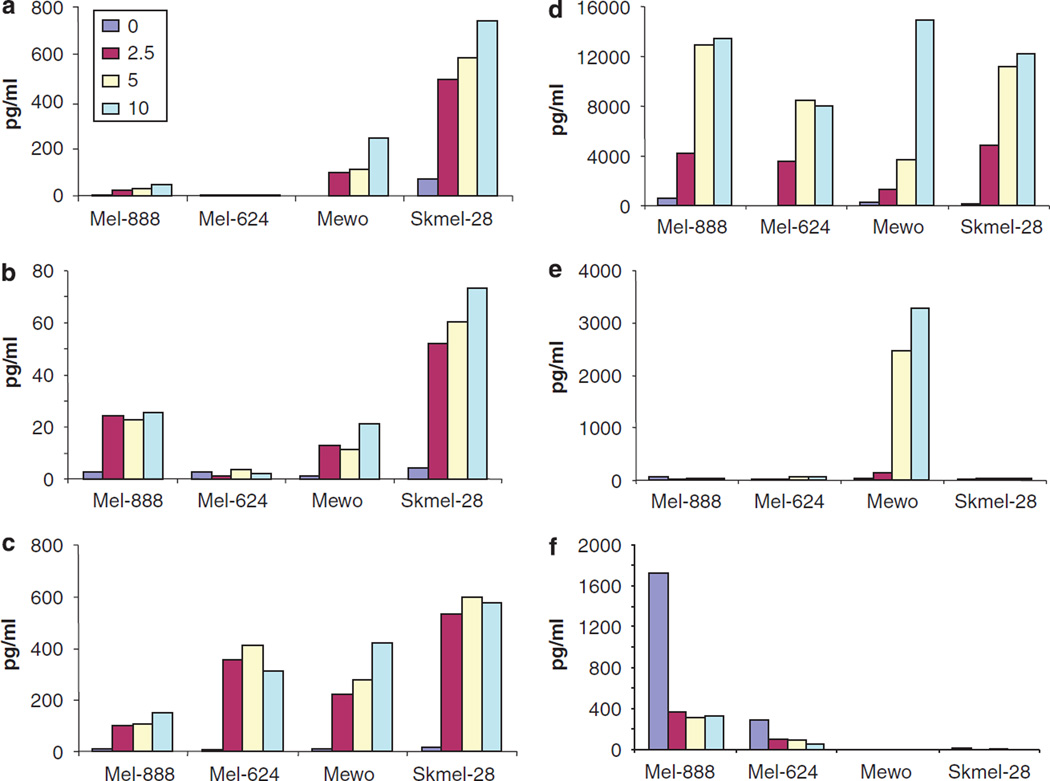
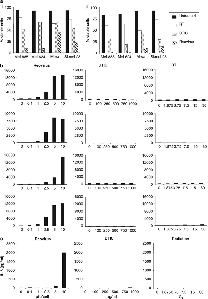
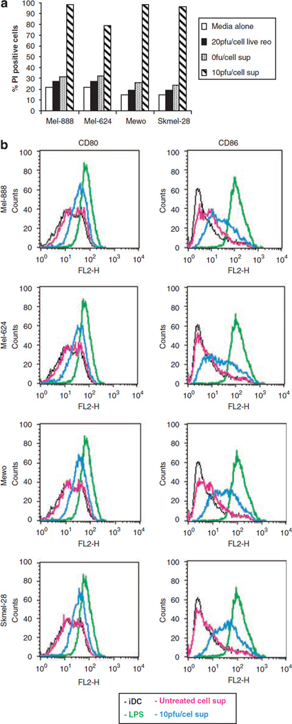
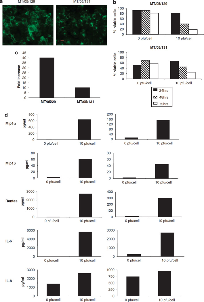
Reovirus is a promising unmodified double-stranded RNA (dsRNA) anti-cancer oncolytic virus, which is thought to specifically target cells with activated Ras. Although reovirus has been tested in a wide range of preclinical models and has entered early clinical trials, it has not previously been tested for the treatment of human melanoma. Here, we show that reovirus effectively kills and replicates in both human melanoma cell lines and freshly resected tumour; intratumoural injection also causes regression of melanoma in a xenograft in vivo model. Reovirus-induced melanoma death is blocked by caspase inhibition and is dependent on constituents of the Ras/RalGEF/p38 pathway. Reovirus melanoma killing is more potent than, and distinct from, chemotherapy or radiotherapy-induced cell death; a range of inflammatory cytokines and chemokines are released by infected tumour cells, while IL-10 secretion is abrogated. Furthermore, the inflammatory response generated by reovirus-infected tumour cells causes bystander toxicity against reovirus-resistant tumour cells and activates human myeloid dendritic cells (DC) in vitro. Hence, reovirus is suitable for clinical testing in melanoma, and may provide a useful danger signal to reverse the immunologically suppressive environment characteristic of this tumour.


Figures

Figure 1
Figure 1
Susceptibility of human melanoma cell lines to reovirus-induced cytotoxicity. (a). Mel-888, Mel-624, Mewo and Skmel-28 cells were either left untreated (control) or treated with 10 PFU/cell reovirus. After 48 or 72 h, all cells were harvested and stained with propidium iodide (PI) before fluorescence-activated cell sorting (FACS) acquisition. Cell death was determined by the proportion of cells staining positive for PI. Average data from four to six independent experiments are shown. Error bars represent s.e.m. (b) Photomicrographs of control (top panel) and 10 PFU/cell reovirus-treated cells (lower panel) were taken 48 h after reovirus infection. (c) Melan-A, Skmel-28, Mel-624, Mel-888, Colo829, A375, WM266 and Mewo cells were treated with serial dilutions of reoviral stock solution (6.9 × 10⁸ PFU/ml) and cell survival was determined by 3-(4,5-dimethylthiazol-2-yl)-2,5-diphenyltetrazolium bromide (MTT) assay. Average data from at least three independent experiments are shown.
Figure 2
Figure 2
Reovirus growth in human melanoma cell lines. (a) Mel-888, Mel-624, Skmel-28, Mewo and Melan-A cells were treated with 10 PFU/cell reovirus for 72 h. Cells and supernatant were harvested, subjected to three rounds of freeze/thaw and the concentration of reovirus determined by plaque assay. The data indicate the fold increase in reovirus compared with input virus and are representative of four independent experiments. (b) To further address the kinetics of viral replication over time, cell lines were treated as in (a), and viral replication determined at 24, 48, 72 and 96 h. (c) Mel-888, Mel-624, Mewo and Skmel-28 cells were either left untreated or treated with 10 PFU/cell reovirus. Whole cell lysates were prepared 0, 3, 8, 16, 24, and 48 h after infection and proteins were separated using SDS-polyacrylamide gel (SDS-PAGE) electrophoresis. Presence of the μ outer capsid protein was determined using a polyclonal antireovirus antibody. β-actin controls demonstrate equal protein loading.
Figure 3
Figure 3
In vivo efficacy of reovirus. Mel-888 xenograft tumours were established in athymic nude mice (eight per group). Tumours were treated with five daily intratumoural injections of either phosphate buffered saline (PBS) or ultraviolet (UV)-inactivated or live reovirus. Tumour growth and survival was monitored up to 70 days after injection. (a) Data represent the percentage of mice surviving over time following injection. (b) Tumour volume of individual mice (each represented by a single line) treated with either PBS or UV-inactivated or live reovirus.
Figure 4
Figure 4
Role of the Ras signalling pathway in reovirus-induced cytotoxicity. Mel-624, Mel-888 and Skmel-28 cells that had been exposed to 10 µm SB202190 (a), U0126 (b) or LY294002 (c) were either left untreated or infected with reovirus at a multiplicity of infection (MOI) approximately equal to the IC50. Cell survival was determined by 3-(4,5-dimethylthiazol-2-yl)-2,5-diphenyltetrazolium bromide (MTT) assay at 96 h. Data are representative of at least three independent experiments. Error bars represent s.e.m.
Figure 5
Figure 5
Mechanism of reovirus-induced cytotoxicity. Mel-888, Mel-624, Mewo and Skmel-28 cells were either left untreated (control), treated with 50 mm ZVAD or 10 PFU/cell reovirus alone or pretreated with 50 µm ZVAD for 1 h before the addition of 10 PFU/cell reovirus. Cells were harvested 48 h after reovirus infection and stained with propidium iodide (PI) before fluorescence-activated cell sorting (FACS) acquisition. Cell death was determined by the proportion of cells staining positive for PI. Data are representative of 2–4 independent experiments.
Figure 6
Figure 6
The inflammatory response associated with reovirus infection. Mel-888, Mel-624, Mewo and Skmel-28 cells were either left untreated or treated with 2.5, 5 or 10 PFU/cell reovirus for 48 h. Cell supernatants were harvested and the production of macrophage inflammatory protein (MIP)-1α (a), MIP-1β (b), RANTES (c), interleukin (IL)-8 (d), IL-6 (e) and IL-10 (f) was determined using ELISA or luminex technology. Data shown are representative of at least two independent experiments.
Figure 7
Figure 7
Reovirus-induced cytotoxicity and proinflammatory response compared with chemotherapy and irradiation. (a) Mel-888, Mel-624, Mewo and Skmel-28 cells were either left untreated or treated with 30 Gy irradiation, 1000 µg/ml dacarbazine (DTIC) or 10 PFU/cell reovirus. Cells were harvested 48 (i) or 72 h (ii) after treatment and stained with propidium iodide (PI) before fluorescence-activated cell sorting (FACS) acquisition. Cell death was determined as the proportion of cells staining positive for PI. (b) Melanoma cell lines were either left untreated, treated with 0.1, 1, 2.5, 5 or 10 PFU/cell reovirus, 100, 250, 500, 750, 1000 µg/ml DTIC or 1.875, 3.75, 7.5, 15 or 30 Gy irradiation. 48 h after treatment, supernatants were harvested and the production of interleukin (IL)-8 was determined by ELISA. (c) Mewo cells were treated as in (b) and the production of IL-6 was determined by ELISA. Data shown are representative of at least two independent experiments.
Figure 8
Figure 8
Functional consequences of reovirus-induced inflammatory cytokine production. (a) Mel-888, Mel-624, Mewo and Skmel-28 cells were either left untreated or treated with 10 PFU/cell reovirus for 48 h. Tumour conditioned media (TCM) was harvested and added mixed at a 1:1 ratio with fresh media to reovirus resistant Daudi cells. After 120 h, viability of Daudi cells was examined by propidium iodide (PI) staining. Data shown are representative of at least three independent experiments. (b) Human immature myeloid dendritic cells (iDC) were left untreated, treated with lipopolysaccharide (LPS) or cultured in tumour conditioned media (TCM) from Mel-888, Mel-624, Mewo and Skmel-28 cells grown ± 10 PFU/cell reovirus for the previous 48 h and expression of CD80 and CD86 determined by flow cytometry. Data shown are representative of three independent experiments.
Figure 9
Figure 9
Response of primary melanoma cells to reovirus. Primary melanoma cells (MT/05/129 and MT/05/131) were isolated as described in Materials and methods. (a) Expression of MART-1 in MT/05/129 and MT/05/131 cells was examined using immunofluorescence. (b) MT/05/129 and MT/05/131 cells were either left untreated or treated with 10 PFU/cell reovirus for 24, 48 or 72 h. Cells were harvested and stained with propidium iodide (PI) before fluorescence-activated cell sorting (FACS) acquisition. (c) MT/05/129 and MT/05/131 cells were treated with 10 PFU/cell reovirus for 72 h. Cells and supernatant were harvested, subjected to three rounds freeze/thaw and the concentration of reovirus was determined by plaque assay. Data shown represent the fold increase in reovirus compared to input virus and are representative of three independent experiments. (d) Cell supernatants were harvested from control- or reovirus-treated MT/05/129 and MT/05/131 cells 48 h after infection. Concentrations of MIP-1α, MIP-1β, RANTES, interleukin (IL)-6 and IL-8 were determined using luminex technology. Data shown are representative of three independent experiments.

References

    1. Selb B, Weber B. A study of human reovirus IgG and IgA antibodies by ELISA and western blot. J Virol Methods. 1994;47:15–25. - PubMed
    1. Coffey MC, Strong JE, Forsyth PA, Lee PW. Reovirus therapy of tumors with activated Ras pathway. Science. 1998;282:1332–1334. - PubMed
    1. Strong JE, Coffey MC, Tang D, Sabinin P, Lee PW. The molecular basis of viral oncolysis: usurpation of the Ras signaling pathway by reovirus. EMBO J. 1998;17:3351–3362. - PMC - PubMed
    1. Strong JE, Lee PW. The v-erbB oncogene confers enhanced cellular susceptibility to reovirus infection. J Virol. 1996;70:612–616. - PMC - PubMed
    1. Marcato P, Shmulevitz M, Lee PW. Connecting reovirus oncolysis and Ras signaling. Cell Cycle. 2005;4:556–559. - PubMed

MeSH terms