Review
. 2008 May;20 Suppl 1(Suppl 1):39-53.
doi: 10.1111/j.1365-2982.2008.01108.x.

Regulation of smooth muscle excitation and contraction

Affiliations
Review

Regulation of smooth muscle excitation and contraction

K M Sanders. Neurogastroenterol Motil. 2008 May.

Abstract

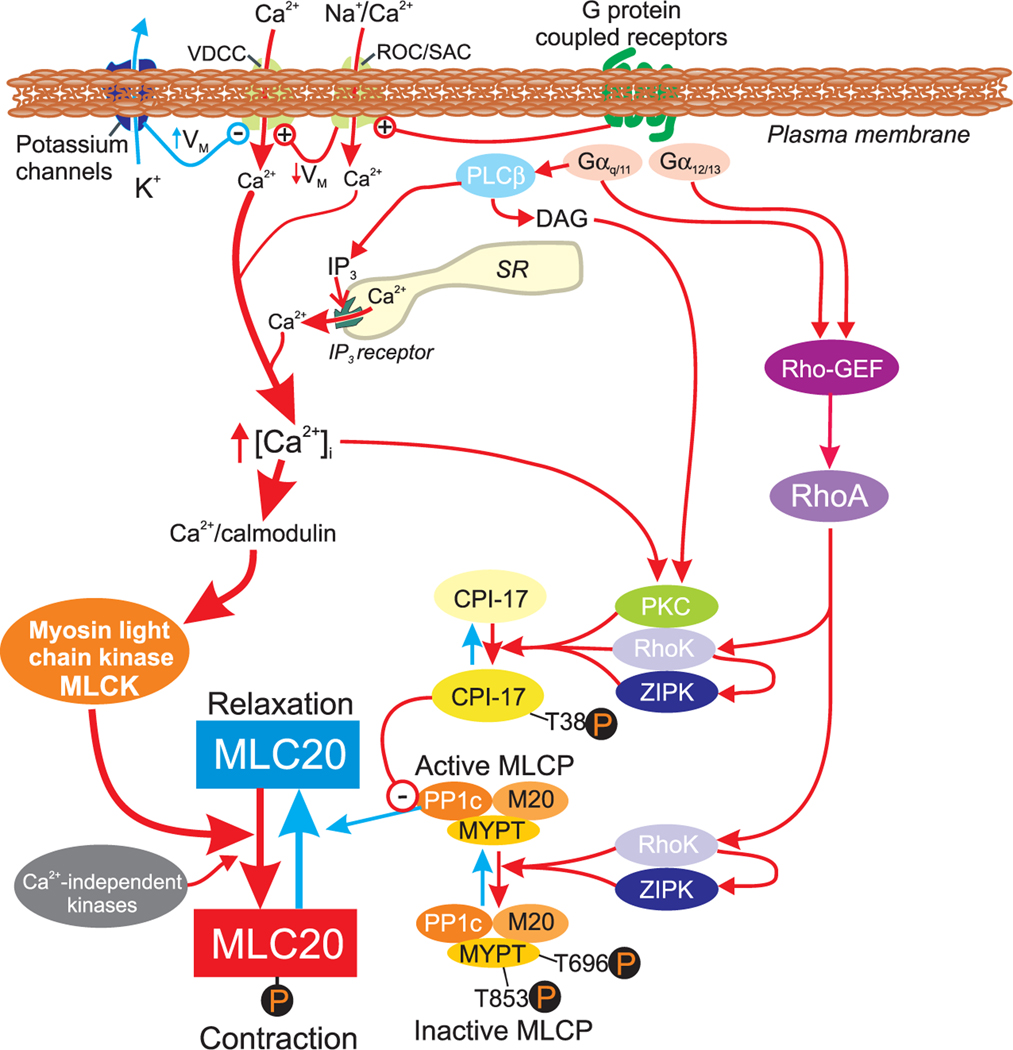
Smooth muscle cells (SMC) make up the muscular portion of the gastrointestinal (GI) tract from the distal oesophagus to the internal anal sphincter. Coordinated contractions of these cells produce the motor patterns of GI motility. Considerable progress was made during the last 20 years to understand the basic mechanisms controlling excitation-contraction (E-C) coupling. The smooth muscle motor is now understood in great molecular detail, and much has been learned about the mechanisms that deliver and recover Ca2+ during contractions. The majority of Ca2+ that initiates contractions comes from the external solution and is supplied by voltage-dependent Ca2+ channels (VDCC). VDCC are regulated largely by the effects of K+ and non-selective cation conductances (NSCC) on cell membrane potential and excitability. Ca2+ entry is supplemented by release of Ca2+ from IP(3) receptor-operated stores and by mechanisms that alter the sensitivity of the contractile apparatus to changes in cytoplasmic Ca2+. Molecular studies of the regulation of smooth muscle have been complicated by the plasticity of SMC and difficulties in culturing these cells without dramatic phenotypic changes. Major questions remain to be resolved regarding the details of E-C coupling in human GI smooth muscles. New discoveries regarding molecular expression that give GI smooth muscle their unique properties, the phenotypic changes that occur in SMC in GI motor disorders, tissue engineering approaches to repair or replace defective muscular regions, and molecular manipulations of GI smooth muscles in animals models and in cell culture will be topics for exciting investigations in the future.


Conflict of interest statement

CONFLICTS OF INTEREST

KMS was supported in this review by grants from the NIDDK: R37 DK40569 and P01 DK41315.

Figures

Figure 1
Figure 1
Schematic representation of major pathways involved in excitation-contraction coupling in gastrointestinal (GI) smooth muscles (Drawing inspired from organization of molecular details in Ref. 94). Most of the Ca2+ required for activation of the contractile apparatus enters cells via VDCC. The major species of VDCC is CaV1.2 channels, but other VDCC are present to a varying extent in GI muscle cells. The open probability of VDCC is regulated negatively by a variety of species of K+ channels expressed by smooth muscle cells. K+ channels are activated by a number of inhibitory agonists (see text for details). The VDCC are regulated positively by receptor-operated (ROC) and stretch-activated (SAC), non-selective cation channels that depolarize smooth muscle cells. ROCs and SACs can also contribute varying amounts to Ca2+ entry, depending upon the molecular species and relative permeability to Na+ and Ca2+. Ca2+ entry can also be supplemented to a varying extent by release of Ca2+ from IP3 receptor-operated Ca2+ channels in the sarcoplasmic reticulum (SR) membrane. IP3 levels are enhanced by synthesis stimulated by agonists binding to G-protein-coupled receptors coupled through Gαq/11 and activation of phospholipase Cβ (PLCβ). A rise in cytoplasmic Ca2+ ([Ca2+]i) binds to the Ca2+ binding protein, calmodulin, and activates MLCK. Myosin light chain kinase phosphorylates the 20 kDa light chain of myosin (MLC20) and facilitates cross-bridge cycling. There are also Ca2+-independent kinases present in smooth muscles that can activate MLCK in a Ca2+-independent manner, but under physiological circumstances, excitation-contraction coupling is largely due to the Ca2+-dependent pathway. Phosphorylation of MLC20 is balanced by MLCP. Dephosphorylation of MLC20 reduces cross-bridge cycling and leads to muscle relaxation. The activity of MLCP is regulated by pathways that in effect regulate the Ca2+ sensitivity of the contractile apparatus. One of these pathways is regulated by G protein regulation of GDP-GTP exchange factor (Rho-GEF), RhoA and activation of RhoK (representing ROCK1 and ROCK2 isoforms). Rho kinase and protein kinase C (PKC) can phosphorylate PKC-potentiated inhibitory protein (CPI-17) at T38 which in turn inhibits the catalytic subunit of MLCP (PP1c). Rho kinase can also phosphorylate the regulatory subunit of MLCP (MYPT) at T696 and T853. Phosphorylation of MYPT decreases the activity of MLCP and its ability to target dephosphorylation of MLC20. Other kinases including zipper-interacting kinase (ZIPK) can also phosphorylate CPI-17 and MYPT. Red arrows depict pathways leading to enhanced contraction; blue arrows indicate pathways leading to reduced contraction.
Figure 2
Figure 2
Contraction of circular and longitudinal gastrointestinal (GI) muscles depends upon Ca2+ entry through VDCC. These traces show contractile responses of the canine gastric antrum, as an example, to carbachol (CCh, 10−7 mol L−1; black bar indicates addition in each panel). Addition of CCh enhanced the amplitude of phasic contraction. Application of CCh within a few minutes after reducing extracellular Ca2+ (to 0.1 mmol L−1) or after addition of nicardipine (10−6 mol L−1) caused much smaller contractile responses. The presence of a phasic contractile response in the presence of nicardipine, albeit of greatly reduced amplitude, probably indicates contributions of other VDCC or ROCs to Ca2+ entry.

Similar articles

Cited by

References

    1. Sanders KM, Koh SD, Ward SM. Interstitial cells of Cajal as pacemakers in the gastrointestinal tract. Annu Rev Physiol 2006; 68: 307–43. - PubMed
    1. Farrugia G. Role of interstitial cells of Cajal in health and disease. Neurogastroenterol Motil 2008; 20 (Suppl. 1): 54–63. - PubMed
    1. Sobue K, Hayashi K, Nishida W. Expressional regulation of smooth muscle cell-specific genes in association with phenotypic modulation. Mol Cell Biochem 1999; 190: 105–18. - PubMed
    1. Arsenian S, Weinhold B, Oelgeschlager M, Ruther U, Nordheim A. Serum response factor is essential for mesoderm formation during mouse embryogenesis. EMBO J 1998; 17: 6289–99. - PMC - PubMed
    1. Angstenberger M, Wegener JW, Pichler BJ et al. Severe intestinal obstruction on induced smooth muscle-specific ablation of the transcription factor SRF in adult mice. Gastroenterology 2007; 133: 1948–59. - PubMed

Publication types