

Vagal and hormonal gut-brain communication: from satiation to satisfaction
- PMID: 18402643
- PMCID: PMC3617963
- DOI: 10.1111/j.1365-2982.2008.01104.x
Vagal and hormonal gut-brain communication: from satiation to satisfaction
Abstract
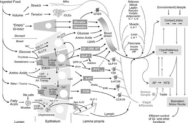
Studying communication between the gut and the brain is as relevant and exciting as it has been since Pavlov's discoveries a century ago. Although the efferent limb of this communication has witnessed significant advances, it is the afferent, or sensory, limb that has recently made for exciting news. It is now clear that signals from the gut are crucial for the control of appetite and the regulation of energy balance, glucose homeostasis, and more. Ghrelin, discovered just a few years ago, is the first gut hormone that increases appetite, and it may be involved in eating disorders. The stable analogue of glucagon-like peptide-1 has rapidly advanced to one of the most promising treatment options for type-2 diabetes. Changes in the signalling patterns of these and other gut hormones best explain the remarkable capacity of gastric bypass surgery to lower food intake and excess body weight. Given the enormous societal implications of the obesity epidemic, these are no small feats. Together with the older gut hormone cholecystokinin and abundant vagal mechanosensors, the gut continuously sends information to the brain regarding the quality and quantity of ingested nutrients, not only important for satiation and meal termination, but also for the appetitive phase of ingestive behaviour and the patterning of meals within given environmental constraints. By acting not only on brainstem and hypothalamus, this stream of sensory information from the gut to the brain is in a position to generate a feeling of satisfaction and happiness as observed after a satiating meal and exploited in vagal afferent stimulation for depression.
Figures
References
-
- Li C, Ford ES, McGuire LC, Mokdad AH. Increasing trends in waist circumference and abdominal obesity among US adults. Obesity (Silver Spring) 2007;15:216–24. - PubMed
-
- Chandrashekar J, Hoon MA, Ryba NJ, Zuker CS. The receptors and cells for mammalian taste. Nature. 2006;444:288–94. - PubMed
-
- Cummings DE. Ghrelin and the short- and long-term regulation of appetite and body weight. Physiol Behav. 2006;89:71–84. - PubMed
-
- Diano S, Farr SA, Benoit SC, et al. Ghrelin controls hippocampal spine synapse density and memory performance. Nat Neurosci. 2006;9:381–8. - PubMed
-
- Kageyama H, Kitamura Y, Hosono T, et al. Visualization of ghrelin-producing neurons in the hypothalamic arcuate nucleus using ghrelin-EGFP transgenic mice. Regul Pept. 2007;145:116–121. - PubMed
Publication types
MeSH terms
Substances
Grants and funding
LinkOut - more resources
Full Text Sources
Other Literature Sources
