. 2008 Apr 10:9:30.
doi: 10.1186/1471-2156-9-30.

GpnmbR150X allele must be present in bone marrow derived cells to mediate DBA/2J glaucoma

Affiliations

GpnmbR150X allele must be present in bone marrow derived cells to mediate DBA/2J glaucoma

Michael G Anderson et al. BMC Genet. .

Abstract

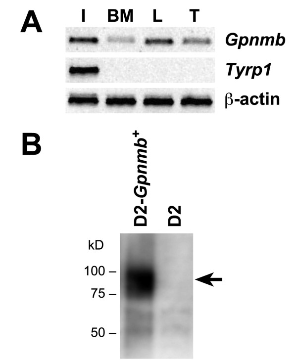
Background: The Gpnmb gene encodes a transmembrane protein whose function(s) remain largely unknown. Here, we assess if a mutant allele of Gpnmb confers susceptibility to glaucoma by altering immune functions. DBA/2J mice have a mutant Gpnmb gene and they develop a form of glaucoma preceded by a pigment dispersing iris disease and abnormalities of the immunosuppressive ocular microenvironment.

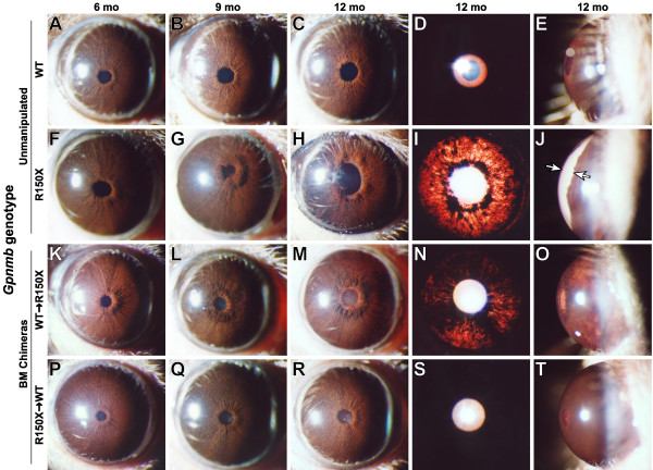
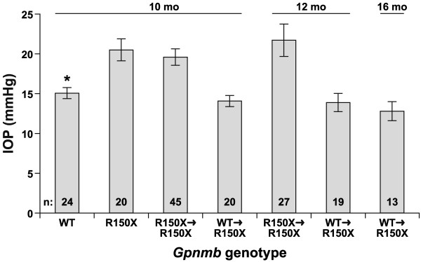
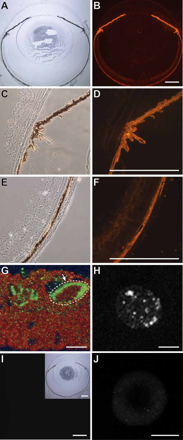
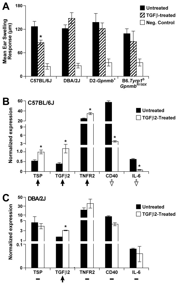
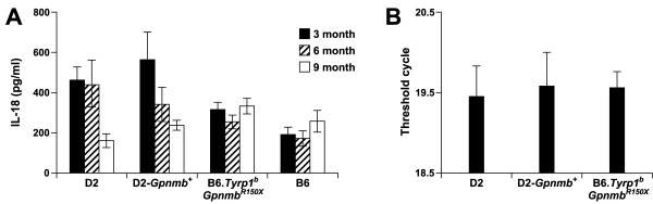
Results: We find that the Gpnmb genotype of bone-marrow derived cell lineages significantly influences the iris disease and the elevation of intraocular pressure. GPNMB localizes to multiple cell types, including pigment producing cells, bone marrow derived F4/80 positive antigen-presenting cells (APCs) of the iris and dendritic cells. We show that APCs of DBA/2J mice fail to induce antigen induced immune deviation (a form of tolerance) when treated with TGFbeta2. This demonstrates that some of the immune abnormalities previously identified in DBA/2J mice result from intrinsic defects in APCs. However, the tested APC defects are not dependent on a mutant Gpnmb gene. Finally, we show that the Gpnmb mediated iris disease does not require elevated IL18 or mature B or T lymphocytes.

Conclusion: These results establish a role for Gpnmb in bone marrow derived lineages. They suggest that affects of Gpnmb on innate immunity influence susceptibility to glaucoma in DBA/2J mice.


Figures

Figure 1
Figure 1
Gpnmb is a candidate for mediating BM derived contributions to D2 glaucoma. A). As expected for a candidate capable of mediating the BM derived contributions to the D2 form of glaucoma, Gpnmb is expressed in BM derived cells. Agarose gel of designated RT-PCR products amplified from iris (I), bone marrow (BM), lymph node (L), or thymus (T) of normal C57BL/6J mice. (B) Iris lysates from D2 and D2-Gpnmb+ mice were analyzed on Western blot with anti-GPNMB antibody (B). There is no protein of the expected size for GPNMB (arrow) in the D2 lysate. The faint bands of lower molecular weight likely represent background cross-reactivity of the antibody.
Figure 2
Figure 2
D2-Gpnmb+ bone marrow suppresses iris depigmentation and anterior chamber enlargement. Representative eyes of the indicated strains and ages are shown. The three left most columns show broad beam illumination to assay for the presence of dispersed pigment within the anterior chamber and iris stromal morphology. The fourth column shows a transilluminating view assaying the degree of iris depigmentation, detectable as red areas within the image where reflected light is passing through the iris. The fifth column show anterior chamber dimensions. Each row represents a different genetic context of either unmanipulated mice (rows 1 & 2) or bone marrow chimeras (rows 3 & 4). (A to E) Unmanipulated D2-Gpnmb+ mice (WT) exhibit a characteristic iris stromal atrophy phenotype caused by the Tyrp1b mutation that is largely devoid of significant pigment dispersion at all examined ages (1–15 mo). Eyes of D2-Gpnmb+ mice do not develop significant transillumination and maintain a normal anterior chamber depth with very little space between the iris and cornea. (F to J) Unmanipulated D2 mice (homozygous for the GpnmbR150X mutation) develop a severe pigment dispersing iris disease. At 5–6 mos, the initial stages of iris disease are apparent by a slight peripupillary thickening and subtle changes to the morphology of the iris stroma (note slightly roughened appearance). Subsequently, dispersed pigment becomes aberrantly deposited on a variety of structures including the surface of the iris, lens, and cornea. Dispersed pigment is also deposited in the aqueous humor drainage structures, leading to increased IOP and enlargement of the anterior chamber (space indicated by arrows in J). (K to O) D2-Gpnmb+marrow transferred to D2.GpnmbR150X recipients (WT → R150X) exhibit a pronounced suppression of the typical D2 disease progression. Most notably, the extent of iris degeneration is lessened (compare the peripupillary region of M vs H, and degree of transillumination defects in N vs I) and the anterior chamber does not become enlarged (compare O vs J). (P to T) D2 marrow transferred to D2-Gpnmb+ recipients (R150X → WT) exhibit no alterations in ocular phenotype compared to unmanipulated D2-Gpnmb+ mice.
Figure 3
Figure 3
Bone marrow genotype affects glaucomatous IOP elevation. Plot of IOP vs Gpnmb genotype, with age and numbers of mice indicated. D2-Gpnmb+ mice exhibit an average IOP typical of many non-glaucomatous mouse strains. As previously described, D2 mice (homozygous for the GpnmbR150X mutation) exhibit an elevated IOP at 10 mo of age, as do D2 mice receiving syngeneic bone marrow transfers (R150X → R150X). Chimeric mice in which D2-Gpnmb+ bone marrow has been transferred into D2 hosts (WT → R150X) continue to maintain non-glaucomatous IOP values, whether examined at 10 mo, or beyond. The IOPs of both Gpnmb+ mice and chimeric mice with Gpnmb+ bone marrow were significantly lower than those of all mice with GpnmbR150X mutant marrow (P < 0.002 for all comparisons at various ages, t test).
Figure 4
Figure 4
GPNMB localizes to multiple pigmented tissues of the eye and ocular APCs. Cryosections of whole eyes from wild-type C57BL/6J mice were imaged in brightfield or phase illumination. GPNMB protein localization was imaged by fluorescent immunohistochemistry and confocal microscopy (red). GPNMB is present in: (A, B) multiple pigmented tissues of the eye, (C, D) iris and pigmented epithelia of the ciliary body, and (E, F) choroid. (G) Iris of normal C57BL/6J mouse showing GPNMB (red) within cells labeled by F4/80 (green). An 8 micron Z-series composite demonstrating F4/80 labeling the border of a single cell (dashed white circle) containing GPNMB. (H) BM derived dendritic cell from D2-Gpnmb+ marrow differentiated in cell culture showing punctate expression of GPNMB, (I, J) Negative controls were labelled and imaged with identical conditions and exhibited negligible signal. (I) Eyes from D2 mice (homozygous for GpnmbR150X) have no signal for GPNMB, an inset on right hand corner shows the brightfield image of the same section. (J) BM derived dendritic cells from D2 mice have no staining for GPNMB. Scale bars A-F, I = 500 μm, G, H, J = 10 μm.
Figure 5
Figure 5
Tolerogenic capacities and expression profiles of Gpnmb deficient APCs. (A) OVA-pulsed APCs isolated from mice of the indicated genotypes were injected into B6D2F1/J recipients. Their ability to induce immune deviation was then measured. Absence of ear swelling indicated functional immune deviation through the suppression of a DTH response to the OVA challenge. We then tested their ability to induce immune deviation as detected by inhibition of DTH i.e. suppression of ear swelling in response to OVA challenge. The experimental summary for each group follows: Untreated (APCs cultured in media, injected into mice, immunization, challenge), TGFβ2-treated (APCs cultured in presence of TGFβ2, injected into mice, immunization, challenge), Neg. Control (challenge only). Induction of immune deviation is detected as the suppression of ear swelling response in the TGFβ2-treated groups as compared to the untreated group. Ear swelling responses are presented as mean ± SEM. (B-C) Gene expression profiles of APCs from indicated genotypes. Real-time PCR was employed to determine the expression levels of the indicated genes and expression of each gene was normalized to Gapdh. To faciliate visualization of the overall pattern of gene expression changes across these genes, the expression changes in response to TGFβ2-treatment are summarized with an arrow (indicating up or down regulation) or a dash (indicating no change). TSP, thrombospondin 1, official gene name Thbs1; TGFβ2, transforming growth factor, beta 2; TNFR2, tumor necrosis factor receptor superfamily, member 1b, official gene name Tnfrsf1b; CD40, CD40 antigen; IL-6, interleukin 6.
Figure 6
Figure 6
IL18 levels do not become elevated in D2 aqueous humor or ciliary body. (A) IL18 levels in aqueous humor of age matched D2, D2-Gpnmb+, B6.Tyrp1bGpnmbR150X, and C57BL/6J mice were determined by ELISA. IL18 levels are expressed as mean +/- SEM from 5 aqueous humor samples in each group. (B) Quantitative RT-PCR analysis comparing expression levels of IL18 transcript in ciliary body enriched dissections of 10.5 mo old D2, D2.Gpnmb+ and B6.Tyrp1bGpnmbR150X mice. The threshold cycle was calculated with normalization to Rn18s and values are expressed as mean +/- SEM, with 3–5 samples in each group.
Figure 7
Figure 7
Loss of adaptive immune reactions has no influence on Gpnmb mediated iris disease. The Rag1tm1Mom mutation acts recessively to result in a loss of adaptive immune responses due to lack of mature B and T lymphocytes. Stocks carrying this mutation were bred to B6.Tyrp1bGpnmbR150X mice. Triple mutant mice were isolated and aged, with typical eyes of indicated genotypes shown. The analysis strikingly demonstrates that the immune deficiency associated with Rag1 mutation has no effect on Gpnmb mediated iris disease. (A) Eyes of young B6 mice with wild-type Tyrp1, Gpnmb, and Rag1 alleles have healthy irides. (B) At 15 mo, mice mutant for Tyrp1 and Gpnmb exhibit iris disease characterized by significant pigment dispersion and iris atrophy. n = 10 eyes, age range 14–16 mo.(C) At 15 mo, immune deficient mice mutant for Tyrp1, Gpnmb, and Rag1 exhibit an iris disease indistinguishable from immune competent mice on the same genetic background. N = 26 eyes.

References

    1. Quigley HA, Broman AT. The number of people with glaucoma worldwide in 2010 and 2020. Br J Ophthalmol. 2006;90:262–267. doi: 10.1136/bjo.2005.081224. - DOI - PMC - PubMed
    1. Allingham RR, Damji KF, Freedman S, Moroi SE, Shafranov G, Shields MB. Shields' Textbook of Glaucoma. 5. Philadelphia: Lippincott Williams and Wilkins; 2004.
    1. Nickells RW, Jampel HD, Zack DJ. Glaucoma. In: Rimoin DL, Conner MJ, Pyeritz RE, Korf BR, editor. Emery & Rimoins Principles and Practices of Medical Genetics. 4. Vol. 3. Churchill Livingstone; 2002. pp. 3491–3512.
    1. Gordon MO, Beiser JA, Brandt JD, Heuer DK, Higginbotham EJ, Johnson CA, Keltner JL, Miller JP, Parrish RK, 2nd, Wilson MR, Kass MA. The Ocular Hypertension Treatment Study: baseline factors that predict the onset of primary open-angle glaucoma. Arch Ophthalmol. 2002;120:714–720. discussion 829–730. - PubMed
    1. Kass MA, Heuer DK, Higginbotham EJ, Johnson CA, Keltner JL, Miller JP, Parrish RK, 2nd, Wilson MR, Gordon MO. The Ocular Hypertension Treatment Study: a randomized trial determines that topical ocular hypotensive medication delays or prevents the onset of primary open-angle glaucoma. Arch Ophthalmol. 2002;120:701–713. discussion 829–730. - PubMed

Publication types

MeSH terms