. 2008 Apr 22;18(8):553-65.
doi: 10.1016/j.cub.2008.02.075. Epub 2008 Apr 10.

The color-vision circuit in the medulla of Drosophila

Affiliations

The color-vision circuit in the medulla of Drosophila

Javier Morante et al. Curr Biol. .

Abstract

Background: Color vision requires comparison between photoreceptors that are sensitive to different wavelengths of light. In Drosophila, this is achieved by the inner photoreceptors (R7 and R8) that contain different rhodopsins. Two types of comparisons can occur in fly color vision: between the R7 (UV sensitive) and R8 (blue- or green sensitive) photoreceptor cells within one ommatidium (unit eye) or between different ommatidia that contain spectrally distinct inner photoreceptors. Photoreceptors project to the optic lobes: R1-R6, which are involved in motion detection, project to the lamina, whereas R7 and R8 reach deeper in the medulla. This paper analyzes the neural network underlying color vision into the medulla.

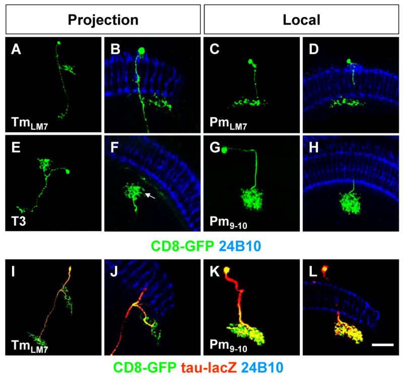
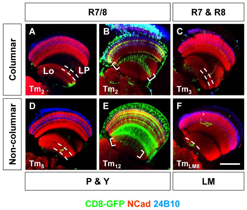
Results: We reconstruct the neural network in the medulla, focusing on neurons likely to be involved in processing color vision. We identify the full complement of neurons in the medulla, including second-order neurons that contact both R7 and R8 from a single ommatidium, or contact R7 and/or R8 from different ommatidia. We also examine third-order neurons and local neurons that likely modulate information from second-order neurons. Finally, we present highly specific tools that will allow us to functionally manipulate the network and test both activity and behavior.

Conclusions: This precise characterization of the medulla circuitry will allow us to understand how color vision is processed in the optic lobe of Drosophila, providing a paradigm for more complex systems in vertebrates.


Figures

Figure 1
Figure 1. Transcription Factor Expression Patterns in the Medulla
(A) Adult optic lobe showing neurons (Elav, blue) in the medulla (Me) cortex (arrows) and medulla rim (arrowheads), lobula (Lo) and lobula plate (LP). (B) High magnification of medulla layers. Brackets show lower medulla layers (M7-M10). (C) Expression pattern of ey-Gal4 driving UAS-CD8::GFP (green). Brackets show lower medulla layers and arrows axonal projections to the lobula. (D) Expression pattern of ap-Gal4. Arrowheads point to L4 neurons and arrows to axonal projections to the lobula. Brackets show medulla layers. (E) Expression pattern of dll-Gal4. (F) Expression pattern of c699-Gal4. Brackets in (E) and (F) show ramifications in the lamina. (G) Non-overlapping expression of ey-Gal4 driving UAS-nuGFP (green), Distal-less antibody (red) and ap-lacZ (blue) in medulla neurons. Inset shows high magnification of medulla cortex. (H) Expression of ey-,dll-,c699-Gal4 driving UAS-nuGFP (green) and ap-lacZ (red) in medulla neurons (blue). Photoreceptor projections visualized with glass-lacZ (green) in (A-B), monoclonal antibody 24B10 (blue) in (C-F) and neuropil stained with DN-Cadherin (red) in (A-F). Scale bar: 50μm (A,C-H) or 15μm (B).
Figure 2
Figure 2. Projection Neurons in the Medulla
MARCM single-cell clones in projection neurons with ap- (A,C,G) or ey-Gal4 (E,I). (A) Tm3-6 cell. Arrow points to ramifications in M7-M9 layers and arrowhead to axonal projection to the lobula. (B) Close-up of Tm3-6 ramifications in photoreceptor layers. (C) Tm2 cell. Arrow points to ramifications in M9 layer and arrowhead to axonal projection to the lobula. (D) Close-up of Tm2 ramifications. Arrowheads points to lateral ramifications and arrow to axonal projection bypassing R7. (E) Non- columnar Tm8 cell. (F) Close-up of Tm8 ramifications. Arrow points to ramifications in M8 layer. (G) A non-columnar TmY2 cell. (H) Close-up of TmY2 ramifications. Arrow points to ramifications in M7 layer. (I) TmP&Y R7 cell. (J) Close-up of TmP&Y R7 ramifications. Arrow points to ramifications in M7 layer. Photoreceptors are visualized with 24B10 (blue). P: pale, Y: yellow. Scale bar: 30μm (A,C,E,G), 15μm (B,D,F,H,J) or 60μm (I).
Figure 3
Figure 3. Microcircuits in the Medulla
MARCM single-cell clones (green) in local and projection neurons using ap-(A,G) or ey-Gal4 (C,E). The comparison between left and right panels highlights the similarity of projection patterns between local and projection neurons that suggests the existence of microcircuits. (A) Mi1 cell. Arrow points to ramifications in M10 layer. (B) Close-up of Mi1. Arrow points to ramification in M3. (C) TmY5a cell. Arrow points to ramifications in lower medulla layers and arrowheads to axonal projections to lobula and lobula plate. (D) Close-up of TmY5a ramifications in M3 and M6 layers (arrowheads). (E-F) Dm1-5 cell. (G) TmY8 cell. Arrowheads point projections to lobula and lobula plate. (H) Close-up of TmY8 ramifications. Photoreceptors are visualized with 24B10 antibody (blue). Scale bar: 15μm (A,B,D,E,F,H) or 40μm (C,G).
Figure 4
Figure 4. Axons in Projection and Local Neurons
Expression of axonal marker UAS-tau-lacZ (red) in ey- (A,F) and ap-Gal4 (C,D) MARCM single-cell clones (green) in projection and local neurons. Axons in TmY5a (A) and Tm7 (D) projection neurons. Inset in (D) shows tau-LacZ staining in axonal projection of Tm7 cell. Close-up of TmY5a (B) and Tm7 (E) ramifications in photoreceptor layers. Axons in Mi9 (C) and Dm1-5 (F) local neurons. Axons are visualized with β-Gal (red) and photoreceptors with 24B10 (blue). Scale bar: 30μm (A,D) or 15μm (B,C,E,F).
Figure 5
Figure 5. Dendrites and Presynaptic Sites in Projection and Local Neurons
Markers driven by specific Gal4 enhancer trap lines indicate the polarity of neurons in the medulla. Co-expression of dendritic UAS-Dscam 17.1 GFP (red) and UAS-AUG::DsRed (green) in (A-B,E-F,I-J), or presynaptic marker UAS-Syt-HA (red) and UAS-CD8:GFP (green) in (C-D,G-H,K-L) in Tm12. (A-D) projection neurons, and Dm6 (E-H) and Pm1 (I-L) local neurons. Close-up of Tm12 (B,D), Dm6 (F,H), and Pm1 (J,L) ramifications (brackets). Arrows in (A,C) point to Syt-positive (C) axonal projections to the lobula and negative for Dscam 17.1 (A). Arrows in (B) point to postsynaptic sites and in (D) to presynaptic sites in lower medulla layers. Photoreceptors visualized with 24B10 (blue). Scale bar: 50μm (A,C,E,G,I,K) or 20μm (B,D,F,H,J,L).
Figure 6
Figure 6. Third Order Neurons: Role of Lower Medulla Layers
MARCM single-cell clones (green) using ey- (A,C,G,I,K) or ap-Gal4 (E) in projection (A,E,I) and local (C,G,K) neurons. (A) TmLM7 cell. (B) Close-up of TmLM7 ramifications. (C) PmLM7 cell. (D) Close-up of PmLM7 ramifications. (E) T3 cell. (F) Close-up of T3 cell ramifications. (G) Pm9-10 cell. (H) Close-up of Pm9-10 ramifications. Co-expression of axonal marker UAS-tau-lacZ (red) in ey-Gal4 (I,K) MARCM-GFP single-cell clones (green) in projection (I) and local neurons (K). (I) Axons in TmLM7 neurons. (J) Close-up of TmLM7 ramifications. (K-L) Axons in Pm9-10 neurons. Axons visualized with β-Gal (red) and photoreceptors with 24B10 (blue). Scale bar: 30μm (A,E,I) or 15μm (B,C,D,F,G,H,J, K,L).
Figure 7
Figure 7. Axonal Projections to the Lobula
Axonal innervation (green) of Tm2 (A), Tm3 (C), Tm5 (D) and TmLM8 (F) MARCM single-cell clones to the lobula (between dashed lines). Expression pattern of otd-Gal4 (B) driving UAS-CD8::GFP (green) in Tm2 cells and photoreceptors, or 2135a-Gal4 in Tm12 cells (E). Brackets in (B,E) point to axonal projections in the lobula. Photoreceptors are visualized with 24B10 (blue) and neuropil with DN-Cadherin (red). Lo: lobula, LM: lower medulla, LP: lobula plate, P: pale, Y: yellow. Scale bar: 50μm.

Comment in

References

    1. Ramon y Cajal S. Histology of the Nervous System of Man and Vertebrates. New York: Oxford University Press; 1995.
    1. Wassle H. Parallel processing in the mammalian retina. Nat Rev Neurosci. 2004;5:747–757. - PubMed
    1. Wolff T, Ready DF. Pattern formation in the Drosophila Retina. In: Bate M, MA A, editors. The Development of Drosophila melanogaster. Vol. 2. Cold Spring Harbor: Cold Spring Harbor Laboratory Press; 1993. pp. 1277–1325.
    1. Hardie R. Functional organization of the fly retina. Vol. 5. Berlin Heidelberg New York: Springer; 1985.
    1. Tang S, Guo A. Choice behavior of Drosophila facing contradictory visual cues. Science. 2001;294:1543–1547. - PubMed

Publication types

Substances