. 2008 Jul;49(7):1409-19.
doi: 10.1194/jlr.M700537-JLR200. Epub 2008 Apr 9.

Intracellular cholesterol transporter StarD4 binds free cholesterol and increases cholesteryl ester formation

Affiliations

Intracellular cholesterol transporter StarD4 binds free cholesterol and increases cholesteryl ester formation

Daniel Rodriguez-Agudo et al. J Lipid Res. 2008 Jul.

Abstract

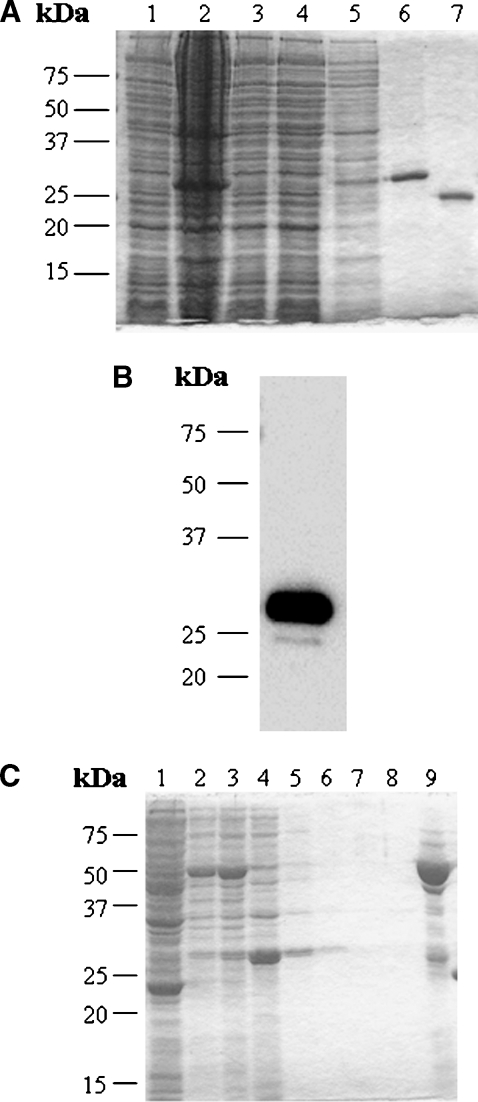
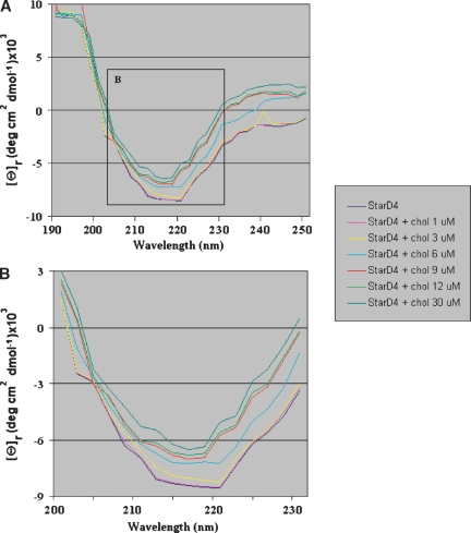
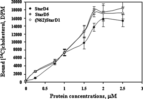
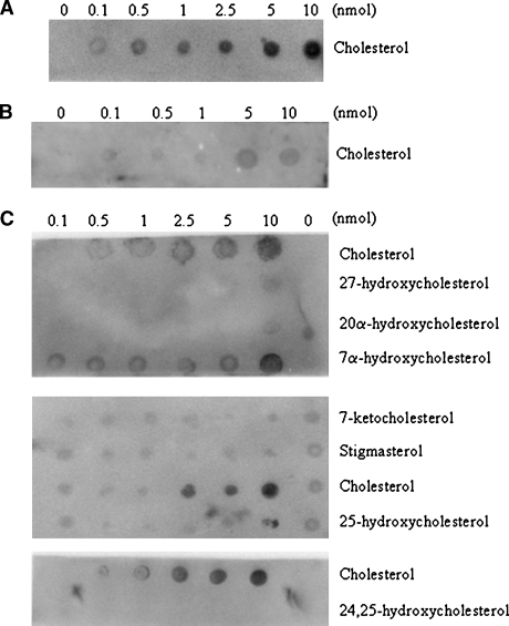
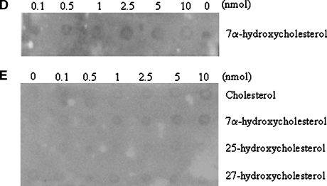
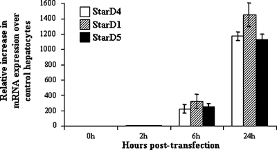
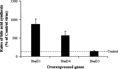
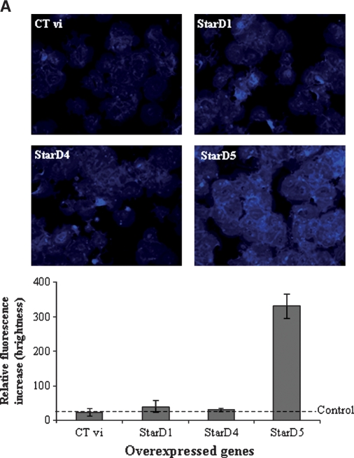
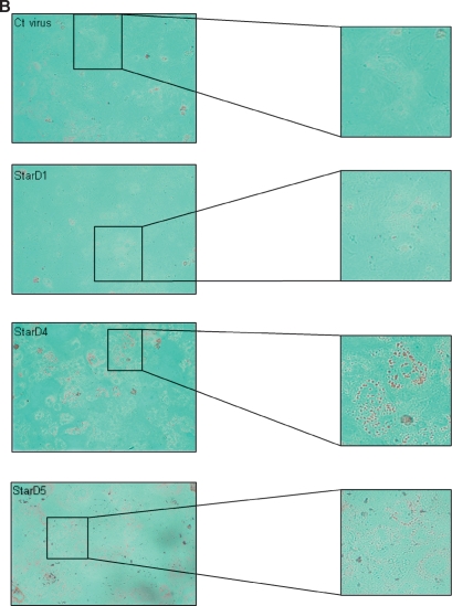
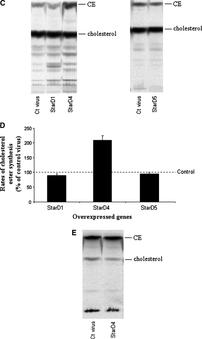
StarD4 protein is a member of the StarD4 subfamily of steroidogenic acute regulatory-related lipid transfer (START) domain proteins that includes StarD5 and StarD6, proteins whose functions remain poorly defined. The objective of this study was to isolate and characterize StarD4's sterol binding and to determine in a hepatocyte culture model its sterol transport capabilities. Utilizing purified full-length StarD4, in vitro binding assays demonstrated a concentration-dependent binding of [(14)C]cholesterol by StarD4 similar to that of the cholesterol binding START domain proteins StarD1 and StarD5. Other tested sterols showed no detectable binding to StarD4, except for 7alpha-hydroxycholesterol, for which StarD4 demonstrated weak binding on lipid protein overlay assays. Subsequently, an isolated mouse hepatocyte model was used to study the ability of StarD4 to bind/mobilize/distribute cellular cholesterol. Increased expression of StarD4 in primary mouse hepatocytes led to a marked increase in the intracellular cholesteryl ester concentration and in the rates of bile acid synthesis. The ability and specificity of StarD4 to bind cholesterol and, as a function of its level of expression, to direct endogenous cellular cholesterol suggest that StarD4 plays an important role as a directional cholesterol transporter in the maintenance of cellular cholesterol homeostasis.


Figures

Fig. 1.
Fig. 1.
Purification and identification of recombinant human StarD4 protein. A: SDS-PAGE analysis and Coomassie blue staining of human StarD4 overexpressed in BL21 cells at each step of the purification. Lane 1, total soluble protein before induction (30 μg); lane 2, total soluble protein after induction (50 μg); lane 3, total soluble protein after incubation with the resin (30 μg); lane 4, proteins eluted with wash buffer I (30 μg); lane 5, proteins eluted with wash buffer II (10 μg); lane 6, protein eluted with lysis buffer plus 1 M imidazole (20 μg); lane 7, StarD4 protein after cleavage of the His tag with recombinant enterokinase. A major protein band was found around 27 kDa. B: Western blot analysis of human StarD4 with monoclonal anti-polyhistidine antibody. Protein eluted with lysis buffer plus 1 M imidazole (200 ng). C: SDS-PAGE analysis and Coomassie blue staining of GST-StarD4 fusion protein overexpressed in BL21 cells at each step of the purification. Lane 1, total soluble protein before induction (50 μg); lane 2, total soluble protein after induction (30 μg); lane 3, total soluble protein before incubation with Glutathione Sepharose 4B (30 μg); lane 4, proteins not bound by the resin (30 μg); lanes 5 to 8, proteins eluted with PBS (10 μg); lane 9, protein eluted with reduced glutathione (20 μg).
Fig. 2.
Fig. 2.
Far-ultraviolet CD spectrum of StarD4. A: The data shown are averages of three scans of the recombinant human protein StarD4 alone at 200 μg/ml (9 μM) and in the presence of cholesterol at different concentrations. B: The inset from A showing a closer look at the spectra between 200 and 230 nm of StarD4 protein alone and in the presence of cholesterol at different concentrations.
Fig. 3.
Fig. 3.
Characterization of StarD4 sterol binding specificities. Different concentrations of His-tagged StarD5, (N62)StarD1 (a completely functional StarD1 protein, as described in Results), and StarD4 were incubated with 1.76 μM [14C]cholesterol for 1 h at 37°C. Data are presented as means ± SEM of three independent experiments.
Fig. 4.
Fig. 4.
Lipid protein overlay (LPO) assays of the steroidogenic acute regulatory-related lipid transfer (START) domain containing protein StarD4 with sterols. The ability of the StarD4 protein to bind various sterols was analyzed. A: Serial dilutions (0–10 nmol) of cholesterol were spotted onto nitrocellulose membranes, which were then incubated with GST-StarD4 protein. B: Binding affinity for cholesterol by GST-StarD4 fusion protein was reduced when the protein was preincubated with cholesterol for 1 h at 37°C previous to the LPO assay. C: Serial dilutions (0–10 nmol) of the indicated sterols were spotted onto nitrocellulose membranes, which were then incubated with GST-StarD4 protein. Cholesterol was included as a positive control in all membranes. D: Binding affinity for 7α-hydroxycholesterol by GST-StarD4 fusion protein was annulled when the protein was preincubated with cholesterol for 1 h at 37°C previous to the LPO assay. E: No nonspecific binding of GST protein was observed when serial dilutions of the indicated sterols were spotted onto nitrocellulose membranes, which were then incubated with GST protein.
Fig. 4.
Fig. 4.
Lipid protein overlay (LPO) assays of the steroidogenic acute regulatory-related lipid transfer (START) domain containing protein StarD4 with sterols. The ability of the StarD4 protein to bind various sterols was analyzed. A: Serial dilutions (0–10 nmol) of cholesterol were spotted onto nitrocellulose membranes, which were then incubated with GST-StarD4 protein. B: Binding affinity for cholesterol by GST-StarD4 fusion protein was reduced when the protein was preincubated with cholesterol for 1 h at 37°C previous to the LPO assay. C: Serial dilutions (0–10 nmol) of the indicated sterols were spotted onto nitrocellulose membranes, which were then incubated with GST-StarD4 protein. Cholesterol was included as a positive control in all membranes. D: Binding affinity for 7α-hydroxycholesterol by GST-StarD4 fusion protein was annulled when the protein was preincubated with cholesterol for 1 h at 37°C previous to the LPO assay. E: No nonspecific binding of GST protein was observed when serial dilutions of the indicated sterols were spotted onto nitrocellulose membranes, which were then incubated with GST protein.
Fig. 5.
Fig. 5.
Quantitative real-time PCR of StarD4, StarD1, and StarD5 mRNA in primary mouse hepatocytes. Following a 2 h transfection with Ad-CMV StarD4, StarD1, or StarD5, levels of the mRNAs were increased in a time-dependent manner over control Ad-CMV transfected hepatocytes. Data are presented as percentage increase over controls (means ± SEM of three independent experiments).
Fig. 6.
Fig. 6.
Effects of StarD1, StarD4, and StarD5 overexpression on the rates of bile acid synthesis in primary mouse hepatocytes. StarD1 and StarD4 overexpression led to an increase in the rates of bile acid synthesis, while StarD5 overexpression did not affect the rates of bile acid synthesis. Bile acid synthesis levels are expressed as percentages of Ad-CMV control transfected cells. Data are presented as means ± SEM of three experiments.
Fig. 7.
Fig. 7.
Effects of the overexpression of different StAR proteins on the levels of free cholesterol and cholesteryl esters in primary mouse hepatocytes. A: Filipin staining for localization of free cholesterol in mouse hepatocytes. Overexpression of StarD1 and StarD4 did not affect the intracellular levels of free cholesterol over control virus, while overexpression of StarD5 increased the levels of free cholesterol up to 12-fold. B: Oil Red O staining of primary mouse hepatocytes overexpressing StarD1, StarD4, StarD5, or control virus. StarD4-overexpressing cells showed a noticeable increase in Oil Red O staining of neutral lipids. C: TLC analysis of sterols in primary mouse hepatocytes after overexpression of StarD1, StarD4, or StarD5. Overexpression of StarD4 in primary hepatocytes led to an increase in cholesteryl ester (CE) levels when medium was supplemented with [14C]cholesterol. No detectable differences were observed in free cholesterol levels following StarD4 overexpression. D: StarD4 overexpression led to increased cholesteryl ester formation, while StarD1 or StarD5 overexpression did not affect cholesteryl ester formation rates. Cholesteryl ester levels are expressed as percentages of Ad-CMV control virus. E: TLC analysis of sterols in primary mouse hepatocytes supplemented with [1-14C]acetate (as substrate for cholesterol synthesis) after overexpression of control StarD4. Overexpression of StarD4 in primary hepatocytes did not increase cholesteryl ester levels from newly synthesized cholesterol. Data are presented as means ± SEM of three independent experiments.
Fig. 7.
Fig. 7.
Effects of the overexpression of different StAR proteins on the levels of free cholesterol and cholesteryl esters in primary mouse hepatocytes. A: Filipin staining for localization of free cholesterol in mouse hepatocytes. Overexpression of StarD1 and StarD4 did not affect the intracellular levels of free cholesterol over control virus, while overexpression of StarD5 increased the levels of free cholesterol up to 12-fold. B: Oil Red O staining of primary mouse hepatocytes overexpressing StarD1, StarD4, StarD5, or control virus. StarD4-overexpressing cells showed a noticeable increase in Oil Red O staining of neutral lipids. C: TLC analysis of sterols in primary mouse hepatocytes after overexpression of StarD1, StarD4, or StarD5. Overexpression of StarD4 in primary hepatocytes led to an increase in cholesteryl ester (CE) levels when medium was supplemented with [14C]cholesterol. No detectable differences were observed in free cholesterol levels following StarD4 overexpression. D: StarD4 overexpression led to increased cholesteryl ester formation, while StarD1 or StarD5 overexpression did not affect cholesteryl ester formation rates. Cholesteryl ester levels are expressed as percentages of Ad-CMV control virus. E: TLC analysis of sterols in primary mouse hepatocytes supplemented with [1-14C]acetate (as substrate for cholesterol synthesis) after overexpression of control StarD4. Overexpression of StarD4 in primary hepatocytes did not increase cholesteryl ester levels from newly synthesized cholesterol. Data are presented as means ± SEM of three independent experiments.

Similar articles

Cited by

References

    1. Hylemon, P. B., W. M. Pandak, and Z. R. Vlahcevic. 2001. The liver: biology and pathobiology. In The Liver. I. M. Arias, J. L. Boyer, F. V. Chisari, et al., editors. Lippincott Williams & Wilkins, Philadelphia, PA. 231–247.
    1. Strauss J. F., III, T. Kishida, L. K. Christenson, T. Fujimoto, and H. Hiroi. 2003. START domain proteins and the intracellular trafficking of cholesterol in steroidogenic cells. Mol. Cell. Endocrinol. 202 59–65. - PubMed
    1. Ponting C. P., and L. Aravind. 2004. START: a lipid-binding domain in StAR, HD-ZIP and signalling proteins. Trends Biochem. Sci. 24 130–132. - PubMed
    1. Rodriguez-Agudo D., S. Ren, P. Hylemon, K. Redford, R. Natarajan, A. Del Castillo, G. Gil, and W. Pandak. 2005. Human StarD5, a cytosolic StAR-related lipid binding protein. J. Lipid Res. 46 1615–1623. - PubMed
    1. Iyer L. M., E. V. Koonin, and L. Aravind. 2001. Adaptations of the helix-grip fold for ligand binding and catalysis in the START domain superfamily. Proteins. 43 134–144. - PubMed

Publication types

MeSH terms