

Epithelial progenitor cells of the embryonic lung and the role of microRNAs in their proliferation
- PMID: 18403323
- PMCID: PMC2645240
- DOI: 10.1513/pats.200710-162DR
Epithelial progenitor cells of the embryonic lung and the role of microRNAs in their proliferation
Abstract
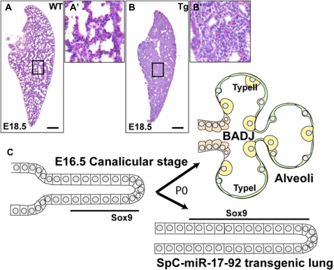
The entire epithelium of the lung is generated from a small pool of undifferentiated progenitor cells. At least during the early stages of development these reside in the distal tips of the embryonic lung. They respond to multiple signals from the surrounding mesenchyme and play a critical role as morphogenetic organizing centers. In addition, they proliferate rapidly and give rise to daughter cells that differentiate into all the specialized epithelial cells types of the newborn lung. Despite the importance of the progenitor cells, we still know relatively little about the mechanisms controlling their proliferation, morphogenesis, and developmental fate. Here, we discuss new data on the potential role of microRNAs in co-coordinately regulating multiple signaling pathways in embryonic progenitor cells. In particular, our recent transgenic experiments suggest that microRNAs encoded by the miR-17-92 cluster positively promote proliferation of epithelial progenitor cells and inhibit their differentiation. We speculate on how this information might be exploited therapeutically in the long term.
Figures

Similar articles
-
Transgenic over-expression of the microRNA miR-17-92 cluster promotes proliferation and inhibits differentiation of lung epithelial progenitor cells.Dev Biol. 2007 Oct 15;310(2):442-53. doi: 10.1016/j.ydbio.2007.08.007. Epub 2007 Aug 9. Dev Biol. 2007. PMID: 17765889 Free PMC article.
-
FGF10 maintains distal lung bud epithelium and excessive signaling leads to progenitor state arrest, distalization, and goblet cell metaplasia.BMC Dev Biol. 2008 Jan 10;8:2. doi: 10.1186/1471-213X-8-2. BMC Dev Biol. 2008. PMID: 18186922 Free PMC article.
-
Human embryonic lung epithelial tips are multipotent progenitors that can be expanded in vitro as long-term self-renewing organoids.Elife. 2017 Jun 30;6:e26575. doi: 10.7554/eLife.26575. Elife. 2017. PMID: 28665271 Free PMC article.
-
Role of microRNAs in stem/progenitor cells and cardiovascular repair.Cardiovasc Res. 2012 Mar 15;93(4):614-22. doi: 10.1093/cvr/cvr311. Epub 2011 Dec 1. Cardiovasc Res. 2012. PMID: 22135162 Review.
-
Stem/progenitor cells in lung development, injury repair, and regeneration.Proc Am Thorac Soc. 2008 Aug 15;5(6):703-6. doi: 10.1513/pats.200801-012AW. Proc Am Thorac Soc. 2008. PMID: 18684721 Free PMC article. Review.
Cited by
-
miR-584 and miR-146 are candidate biomarkers for acute respiratory distress syndrome.Exp Ther Med. 2021 May;21(5):445. doi: 10.3892/etm.2021.9873. Epub 2021 Mar 1. Exp Ther Med. 2021. PMID: 33747181 Free PMC article.
-
Eya1 controls cell polarity, spindle orientation, cell fate and Notch signaling in distal embryonic lung epithelium.Development. 2011 Apr;138(7):1395-407. doi: 10.1242/dev.058479. Development. 2011. Retraction in: Development. 2017 Oct 15;144(20):3849. doi: 10.1242/dev.159673. PMID: 21385765 Free PMC article. Retracted.
-
Combined KIT and FGFR2b signaling regulates epithelial progenitor expansion during organogenesis.Stem Cell Reports. 2013 Dec 12;1(6):604-19. doi: 10.1016/j.stemcr.2013.10.013. eCollection 2013. Stem Cell Reports. 2013. PMID: 24371813 Free PMC article.
-
MicroRNA-127 modulates fetal lung development.Physiol Genomics. 2009 May 13;37(3):268-78. doi: 10.1152/physiolgenomics.90268.2008. Epub 2009 Mar 17. Physiol Genomics. 2009. PMID: 19439715 Free PMC article.
-
Prenatal retinoid deficiency leads to airway hyperresponsiveness in adult mice.J Clin Invest. 2014 Feb;124(2):801-11. doi: 10.1172/JCI70291. Epub 2014 Jan 9. J Clin Invest. 2014. PMID: 24401276 Free PMC article.
References
-
- Bushati N, Cohen SM. MicroRNA functions. Annu Rev Cell Dev Biol 2007;23:175–205. - PubMed
-
- Lim LP, Glasner ME, Yekta S, Burge CB, Bartel DP. Vertebrate microRNA genes. Science 2003;299:1540. - PubMed
-
- Valencia-Sanchez MA, Liu J, Hannon GJ, Parker R. Control of translation and mRNA degradation by miRNAs and siRNAs. Genes Dev 2006;20:515–524. - PubMed
-
- He L, Hannon GJ. MicroRNAs: small RNAs with a big role in gene regulation. Nature Rev 2004;5:522–531. - PubMed
Publication types
MeSH terms
Substances
Grants and funding
LinkOut - more resources
Full Text Sources
Other Literature Sources
Medical