. 2008 May;36(9):3101-17.
doi: 10.1093/nar/gkn162. Epub 2008 Apr 10.

The three SoxC proteins--Sox4, Sox11 and Sox12--exhibit overlapping expression patterns and molecular properties

Affiliations

The three SoxC proteins--Sox4, Sox11 and Sox12--exhibit overlapping expression patterns and molecular properties

Peter Dy et al. Nucleic Acids Res. 2008 May.

Abstract

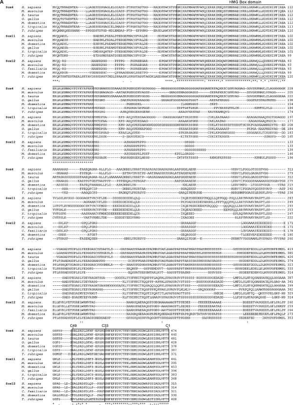
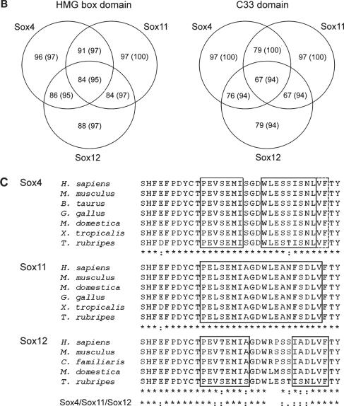
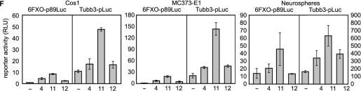
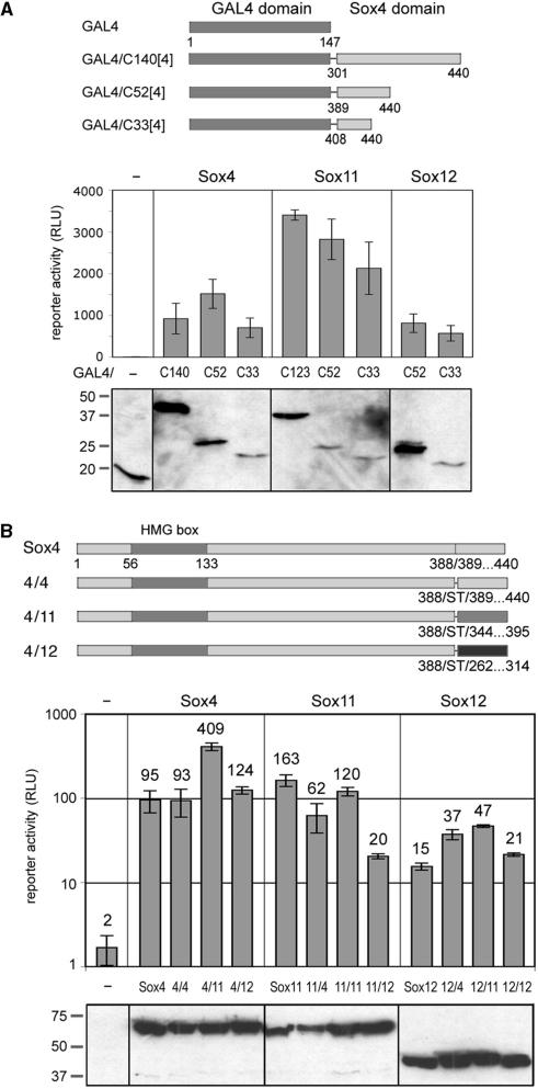
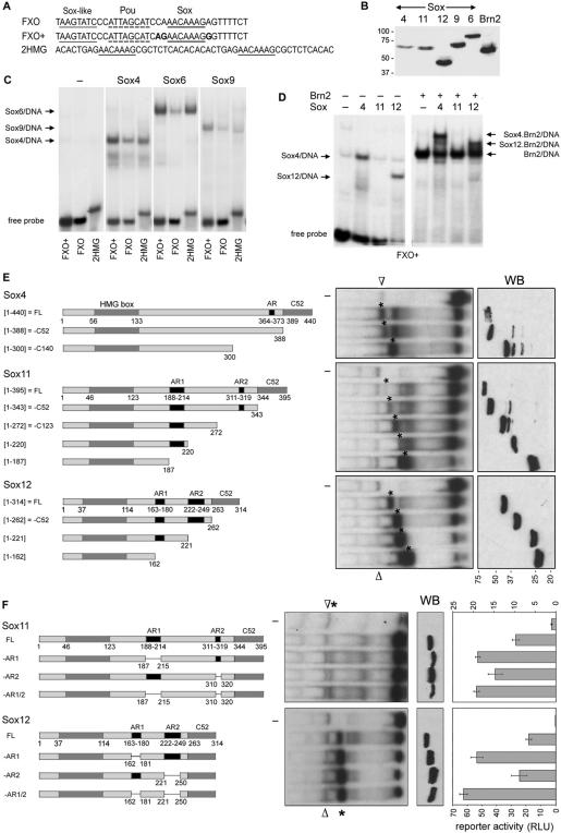
The group C of Sry-related high-mobility group (HMG) box (Sox) transcription factors has three members in most vertebrates: Sox4, Sox11 and Sox12. Sox4 and Sox11 have key roles in cardiac, neuronal and other major developmental processes, but their molecular roles in many lineages and the roles of Sox12 remain largely unknown. We show here that the three genes are co-expressed at high levels in neuronal and mesenchymal tissues in the developing mouse, and at variable relative levels in many other tissues. The three proteins have conserved remarkable identity through evolution in the HMG box DNA-binding domain and in the C-terminal 33 residues, and we demonstrate that the latter residues constitute their transactivation domain (TAD). Sox11 activates transcription several times more efficiently than Sox4 and up to one order of magnitude more efficiently than Sox12, owing to a more stable alpha-helical structure of its TAD. This domain and acidic domains interfere with DNA binding, Sox11 being most affected and Sox4 least affected. The proteins are nevertheless capable of competing with one another in reporter gene transactivation. We conclude that the three SoxC proteins have conserved overlapping expression patterns and molecular properties, and might therefore act in concert to fulfill essential roles in vivo.


Figures

Figure 1.
Figure 1.
Comparison of the SoxC gene expression pattern in the mouse. (A) Northern blots of total RNA from E12.5 mouse embryos hybridized with Sox4, Sox11 and Sox12 probes, as indicated. A picture of the RNA stained with ethidium bromide before blotting is shown in the left lane. The migration level and size of DNA markers run on the same gel are indicated on the right. The three probes had the same labeling specificity and the three blots were exposed for the length of time to allow direct comparison of RNA levels. (B) Northern blots showing the relative expression levels of the SoxC RNAs in various tissues of 2-day-old mice and in cultured cells. WE, whole embryo at E12.5; Br, brain; He, heart; Lu, lung; Li, liver; Pa, pancreas; SI, small intestine; Nph, mouse embryo primary neurospheres; Cos1, monkey kidney fibroblastic cell line; MC3T3, preosteoblastic MC3T3-E1 cell line. The three probes had the same labeling specificity and the blots in each of the three sub-panels were exposed for the same length of time to allow direct comparison of RNA levels. The size of the Sox11 RNA in monkey Cos1 cells was ∼5 kb, instead of ∼7 kb in mouse tissues and cells (unpublished results). (C) RNA in situ hybridization of adjacent mid-sagittal sections of E12.5 mouse embryos with Sox4, Sox11 and Sox12 probes. The left-side section was stained with hematoxylin and eosin (H&E). CM, craniofacial (skeletogenic) mesenchyme; CNS, central nervous system; GM, genital tubercle mesenchyme; H, heart; TO, tongue; VM, vertebral column (skeletogenic) mesenchyme. (D) RNA in situ hybridization of adjacent coronal sections of mouse embryo heads at E14.5 and E18.5 with SoxC probes. EL, eyelid mesenchyme; J, presumptive jaw mesenchyme; M, mesenchyme; OE, olfactory epithelium; PS, palatal shelves; R, retina; S, skin primordium TO, tongue; TE, teeth. (E) RNA in situ hybridization of various developing organs in E14.5 to E18.5 embryos with SoxC probes, as indicated. Ep, epithelium; HF, hair follicle; Mg, midgut; Ms, mesenchyme; Sp, spleen; Th, thymus.
Figure 2.
Figure 2.
Comparison of the SoxC protein sequences in vertebrates. (A) ClustalX alignment of the Sox4, Sox11 and Sox12 protein sequences in various vertebrate species. Symbols underneath the alignments denote fully conserved amino acid residues (asterisk), conservative changes (colon) and semi-conservative changes (dot). Boxes highlight the highly conserved HMG box domain and the C-terminal domain (from residues C49 and C33 to C1), and as well as partially conserved acidic, serine-rich and glycine-rich domains identified by the ScanProsite tool. (B) Venn diagrams depicting the degrees of identity and similarity (the latter in parentheses) of SoxC orthologues in the HMG box and C33 domains. Each of the Sox4, Sox11 and Sox12 subgroups of orthologues is schematized as a circle. Numbers indicate the percentage of conservation between orthologues in the same subgroup (nonoverlapping circle areas) and between orthologues in different groups (overlapping circle areas). (C) ClustalX alignment of SoxC C33 domain sequences. Boxes with a continuous line highlight residues in α-helical conformation and the box with a dotted line highlights residues in extended conformation.
Figure 2.
Figure 2.
Comparison of the SoxC protein sequences in vertebrates. (A) ClustalX alignment of the Sox4, Sox11 and Sox12 protein sequences in various vertebrate species. Symbols underneath the alignments denote fully conserved amino acid residues (asterisk), conservative changes (colon) and semi-conservative changes (dot). Boxes highlight the highly conserved HMG box domain and the C-terminal domain (from residues C49 and C33 to C1), and as well as partially conserved acidic, serine-rich and glycine-rich domains identified by the ScanProsite tool. (B) Venn diagrams depicting the degrees of identity and similarity (the latter in parentheses) of SoxC orthologues in the HMG box and C33 domains. Each of the Sox4, Sox11 and Sox12 subgroups of orthologues is schematized as a circle. Numbers indicate the percentage of conservation between orthologues in the same subgroup (nonoverlapping circle areas) and between orthologues in different groups (overlapping circle areas). (C) ClustalX alignment of SoxC C33 domain sequences. Boxes with a continuous line highlight residues in α-helical conformation and the box with a dotted line highlights residues in extended conformation.
Figure 3.
Figure 3.
Comparison of the transactivation activity of the mouse SoxC proteins. (A) Schematic of Sox reporter genes. Each reporter features a minimal Col2a1 promoter (p89) driving the firefly luciferase gene. The FXO, HMG and 48bp Col2a1 intron-1 fragment sequences were cloned as 6, 2 or 4 tandem copies, respectively, directly upstream of the promoter. The Sox recognition sites are underlined with a continuous line. The POU-domain recognition site and a Sox-like site in the FXO sequence are shown with a dotted and a double line, respectively. (B) Comparison of the ability of the SoxC proteins to transactivate 6FXO-p89Luc. The 6FXO-p89Luc was transfected into Cos1 cells with 0, 10, 30, 100 or 300 ng of either SoxC expression plasmid, supplemented with empty expression plasmid up to 300 ng. Reporter activities are plotted against the amount of SoxC expression plasmid. The western blot illustrates the amount of SoxC protein present in the cells at the end of the experiment. The amount of extract loaded on the gel for each condition was normalized for transfection efficiency. An anti-FLAG antibody was used to detect the proteins. This blot demonstrates that all SoxC proteins were produced at a similar level for each expression plasmid amount, and therefore that the differences seen between the SoxC proteins in transactivation efficiency are due to differences in the intrinsic properties of the proteins rather than to differences in their relative amounts. The Mr of protein standards is indicated on the left of the blot. Note that each SoxC protein exhibits an apparent Mr (Sox4, 69k; Sox11, 68k; Sox12, 45k) slightly larger than predicted (Sox4, 45k; Sox11, 43k; Sox12, 34k). (C) Comparison of the ability of the three SoxC proteins to transactivate the 6FXO-p89Luc reporter in synergy with Brn2. Cos1 cells were transfected with the reporters and 100 ng Brn2 and 100 ng of SoxC expression plasmid. Note that the scale of the graph is logarithmic. Reporter activities are indicated. (D) Comparison of the transactivation efficiency of SoxC proteins with and without an N-terminal FLAG epitope. Cos1 cells were transfected with 200 ng expression plasmid for SoxC proteins with (F4, F11 and F12) or without (4, 11 or 12) the FLAG epitope fused at the N-terminus. All SoxC proteins were detected by western blot using a SoxC antibody. (E) Comparison of the ability of the three SoxC proteins and Sox9 to transactivate the 6FXO-p89Luc, 2HMG-p89Luc and 4x48-p89Luc reporters. Cos1 cells were transfected with the reporters and 200 ng of Sox expression plasmid. SoxC proteins were detected with anti-FLAG antibody. Note that it is important to consider the relative amounts of protein expressed in each condition to properly interpret data. (F) Comparison of the ability of the three SoxC proteins to transactivate the 6FXO-p89Luc and Tubb3-pLuc reporters in Cos1 cells, MC3T3-E1 cells and neurospheres. Cells were co-transfected with the reporters and with 200 ng (Cos1 and MC3T3-E1) or 600 ng (neurospheres) of SoxC expression plasmid.
Figure 3.
Figure 3.
Comparison of the transactivation activity of the mouse SoxC proteins. (A) Schematic of Sox reporter genes. Each reporter features a minimal Col2a1 promoter (p89) driving the firefly luciferase gene. The FXO, HMG and 48bp Col2a1 intron-1 fragment sequences were cloned as 6, 2 or 4 tandem copies, respectively, directly upstream of the promoter. The Sox recognition sites are underlined with a continuous line. The POU-domain recognition site and a Sox-like site in the FXO sequence are shown with a dotted and a double line, respectively. (B) Comparison of the ability of the SoxC proteins to transactivate 6FXO-p89Luc. The 6FXO-p89Luc was transfected into Cos1 cells with 0, 10, 30, 100 or 300 ng of either SoxC expression plasmid, supplemented with empty expression plasmid up to 300 ng. Reporter activities are plotted against the amount of SoxC expression plasmid. The western blot illustrates the amount of SoxC protein present in the cells at the end of the experiment. The amount of extract loaded on the gel for each condition was normalized for transfection efficiency. An anti-FLAG antibody was used to detect the proteins. This blot demonstrates that all SoxC proteins were produced at a similar level for each expression plasmid amount, and therefore that the differences seen between the SoxC proteins in transactivation efficiency are due to differences in the intrinsic properties of the proteins rather than to differences in their relative amounts. The Mr of protein standards is indicated on the left of the blot. Note that each SoxC protein exhibits an apparent Mr (Sox4, 69k; Sox11, 68k; Sox12, 45k) slightly larger than predicted (Sox4, 45k; Sox11, 43k; Sox12, 34k). (C) Comparison of the ability of the three SoxC proteins to transactivate the 6FXO-p89Luc reporter in synergy with Brn2. Cos1 cells were transfected with the reporters and 100 ng Brn2 and 100 ng of SoxC expression plasmid. Note that the scale of the graph is logarithmic. Reporter activities are indicated. (D) Comparison of the transactivation efficiency of SoxC proteins with and without an N-terminal FLAG epitope. Cos1 cells were transfected with 200 ng expression plasmid for SoxC proteins with (F4, F11 and F12) or without (4, 11 or 12) the FLAG epitope fused at the N-terminus. All SoxC proteins were detected by western blot using a SoxC antibody. (E) Comparison of the ability of the three SoxC proteins and Sox9 to transactivate the 6FXO-p89Luc, 2HMG-p89Luc and 4x48-p89Luc reporters. Cos1 cells were transfected with the reporters and 200 ng of Sox expression plasmid. SoxC proteins were detected with anti-FLAG antibody. Note that it is important to consider the relative amounts of protein expressed in each condition to properly interpret data. (F) Comparison of the ability of the three SoxC proteins to transactivate the 6FXO-p89Luc and Tubb3-pLuc reporters in Cos1 cells, MC3T3-E1 cells and neurospheres. Cells were co-transfected with the reporters and with 200 ng (Cos1 and MC3T3-E1) or 600 ng (neurospheres) of SoxC expression plasmid.
Figure 4.
Figure 4.
The SoxC C33 domain is required for transactivation. (A) Schematic of SoxC proteins truncated in the C-terminus. The name of each protein is indicated on the left of the rectangle representing it. Numbers designate the first and last protein residues and relevant domain boundaries. A thin line denotes internal deletions. (B) Effect of deleting the C52 or C33 region on the ability of SoxC proteins to transactivate 6FXO-p89Luc. Cos1 cells were transfected with 200 ng expression plasmid encoding a full-length SoxC protein (FL) or a SoxC protein lacking the C52 (−52) or C33 (−33) domain. Reporter activities are indicated. SoxC proteins were detected by western blot with anti-FLAG antibody. Note partial degradation of the Sox11-C33 and Sox11-C52 proteins. (C) Effect of deleting the SoxC C-terminal third or an internal region on the ability of Sox4 and Sox11 to transactivate 6FXO-p89Luc. Cos1 cells were transfected with 200 ng expression plasmid encoding a full-length SoxC protein (FL), a SoxC protein lacking the C-terminal third (−140 and −123) or a Sox protein lacking an internal segment (−140/53 and −123/53). SoxC proteins were detected by western blot with anti-FLAG antibody. (D) Effect of deleting the C33 domain on the ability of SoxC proteins to synergize with Brn2 in transactivating 6FXO-p89Luc. Cos1 cells were transfected with 100 ng expression plasmid encoding Brn2 and 100 ng of expression plasmid encoding a full-length (FL) SoxC protein or a SoxC protein lacking C33 (−C33). The western blot with a FLAG antibody shows the amount of Brn2 protein (arrow) made in each culture and some of the SoxC proteins. The western blot with the SoxC antibody shows the relative amounts of SoxC proteins made in each culture. These proteins appear as doublet, an electrophoresis artifact.
Figure 5.
Figure 5.
The SoxC C33 domain is sufficient for transactivation. (A) Schematic of GAL4-SoxC fusion proteins and transactivation of pG5Luc by these proteins. Cos1 cells were transfected with 100 ng GAL4/SoxC expression plasmid. Reporter activities are presented in comparison with the amount of GAL4/SoxC fusion proteins present in each culture at the end of the experiment and detected with anti-GAL4 antibody. (B) Schematic of Sox4 proteins with swapped SoxC C52 regions and transactivation of 6FXO-p89Luc by these swapped SoxC proteins. The swapped Sox11 and Sox12 proteins were made as shown for Sox4. Cos1 cells were transfected with 100 ng swapped SoxC expression plasmid. SoxC proteins were detected with an anti-FLAG antibody.
Figure 6.
Figure 6.
Comparison of the DNA-binding efficiency of the SoxC proteins. (A) Sequences of FXO, FXO+ and 2HMG EMSA probes. Sox, Sox-like and POU domain protein binding sites are underlined. (B) Western blot with anti-FLAG antibody demonstrating the relative amounts of proteins used in EMSA in (C) and (D). (C) EMSA of Sox4, Sox6 and Sox9 with the FXO, FXO+ and 2HMG probes. The Sox protein/DNA complexes are identified with arrows. (D) EMSA of SoxC proteins with FXO+ in the presence (+) or absence (−) of Brn2. Specific protein/DNA complexes are identified with arrows. Note that the Sox4/DNA complex migrates with the same mobility as a less abundant complex present in all samples and that the free probe ran off the right part of the gel. (E) Effect of deleting an increasing segment of the SoxC C-terminus on the ability of the proteins to bind DNA. SoxC proteins were made in Cos1 cells as schematized and used in EMSA with the FXO+ probe. The SoxC/DNA complexes are identified in the EMSA with asterisks and a nonspecific complex with an open triangle. A western blot (WB) hybridized with an anti-FLAG antibody shows the relative amounts of the various proteins. (F) Effect of deleting the acidic regions of Sox11 and Sox12 on the protein's ability to bind DNA and transactivate. Cos1 cells were transfected with 200 ng of expression plasmid encoding full-length or deletion mutant proteins, as schematized, and with the 6FXO-p89Luc reporter and control plasmids. SoxC proteins were tested in EMSA with the FXO+ probe and detected in western blot with an anti-FLAG antibody.
Figure 7.
Figure 7.
Dominant-negative interference of SoxC proteins truncated in the C-terminus. (A) Test of the ability of SoxC proteins lacking the C33 domain to interfere with the activity of SoxC full-length proteins. Cos1 cells were transfected with 100 ng Brn2 expression plasmid, 100 ng SoxC full-length expression plasmid and 100 ng truncated SoxC protein expression plasmid, and with the 6FXO-p89Luc reporter and control plasmids. SoxC and Brn2 proteins were detected by western blot using an anti-FLAG antibody. (B) Test of the ability of Sox11 proteins lacking various lengths of the C-terminus to interfere with the activity of full-length Sox11. Cos1 cells were transfected with 100 ng Sox11 full-length expression plasmid and either 20 or 100 ng truncated Sox11 expression plasmid, as indicated, and with the 6FXO-p89Luc reporter and control plasmids. The Sox11 proteins were detected by western blot using an anti-FLAG antibody. (C) Inhibition of the activity of the Tubb3 promoter by SoxC proteins lacking the C33 domain. Cos1 cells were transiently transfected with the Tubb3-pLuc reporter and with 600 ng of the expression plasmids for the mutant SoxC proteins.

References

    1. Schepers GE, Teasdale RD, Koopman P. Twenty pairs of Sox: extent, homology, and nomenclature of the mouse and human Sox transcription factor gene families. Dev. Cell. 2002;3:167–170. - PubMed
    1. Lefebvre V, Dumitriu B, Penzo-Méndez A, Han Y, Pallavi B. Control of cell fate and differentiation by Sry-related high-mobility-group box (Sox) transcription factors. Int. J. Biochem. Cell. Biol. 2007;39:2195–2214. - PMC - PubMed
    1. Koopman P, Gubbay J, Vivian N, Goodfellow P, Lovell-Badge R. Male development of chromosomally female mice transgenic for Sry. Nature. 1991;351:117–121. - PubMed
    1. Laudet V, Stehelin D, Clevers H. Ancestry and diversity of the HMG box superfamily. Nucleic Acids Res. 1993;21:2493–2501. - PMC - PubMed
    1. Bianchi ME, Agresti A. HMG proteins: dynamic players in gene regulation and differentiation. Curr. Opin. Genet. Dev. 2005;15:496–506. - PubMed

Publication types

MeSH terms