

The effects of experimental muscle and skin pain on the static stretch sensitivity of human muscle spindles in relaxed leg muscles
- PMID: 18403422
- PMCID: PMC2536575
- DOI: 10.1113/jphysiol.2008.151746
The effects of experimental muscle and skin pain on the static stretch sensitivity of human muscle spindles in relaxed leg muscles
Abstract
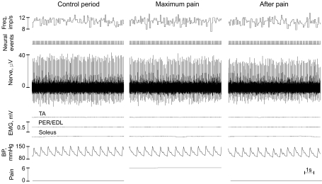
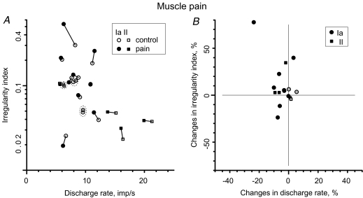
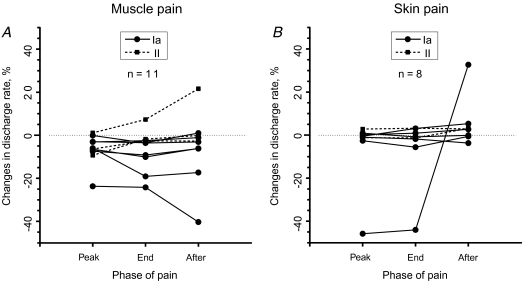
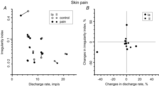
Animal studies have shown that noxious inputs onto gamma-motoneurons can cause an increase in the activity of muscle spindles, and it has been proposed that this causes a fusimotor-driven increase in muscle stiffness that is believed to underlie many chronic pain syndromes. To test whether experimental pain also acts on the fusimotor system in humans, unitary recordings were made from 19 spindle afferents (12 Ia, 7 II) located in the ankle and toe extensors or peronei muscles of awake human subjects. Muscle pain was induced by bolus intramuscular injection of 0.5 ml 5% hypertonic saline into tibialis anterior (TA); skin pain was induced by 0.2 ml injection into the overlying skin. Changes in fusimotor drive to the muscle spindles were inferred from changes in the mean discharge frequency and discharge variability of spindle endings in relaxed muscle. During muscle pain no afferents increased their discharge activity: seven afferents (5 Ia, 2 II) showed a decrease and six (4 Ia, 2 II) afferents were not affected. During skin pain of 13 afferents discharge rate increased in one (Ia) and decreased in two (1 Ia, 1 II). On average, the overall discharge rate decreased during muscle pain by 6.1% (P < 0.05; Wilcoxon), but remained essentially the same during skin pain. There was no detectable correlation between subjective pain level and the small change in discharge rate of muscle spindles. Irrespective of the type of pain, discharge variability parameters were not influenced (P > 0.05; Wilcoxon). We conclude that, contrary to the 'vicious cycle' hypothesis, acute activation of muscle or skin nociceptors does not cause a reflex increase in fusimotor drive in humans. Rather, our results are more aligned with the pain adaptation model, based on clinical studies predicting pain-induced reductions of agonist muscle activity.
Figures
Similar articles
-
Effects of tonic muscle pain on fusimotor control of human muscle spindles during isometric ankle dorsiflexion.J Neurophysiol. 2019 Apr 1;121(4):1143-1149. doi: 10.1152/jn.00862.2018. Epub 2019 Jan 30. J Neurophysiol. 2019. PMID: 30699044
-
Tonic muscle pain does not increase fusimotor drive to human leg muscles: implications for chronic muscle pain.Exp Physiol. 2013 Jun;98(6):1125-32. doi: 10.1113/expphysiol.2012.071670. Epub 2013 Feb 15. Exp Physiol. 2013. PMID: 23417691
-
Fusimotor reflexes in relaxed forearm muscles produced by cutaneous afferents from the human hand.J Physiol. 1994 Sep 15;479 ( Pt 3)(Pt 3):499-508. doi: 10.1113/jphysiol.1994.sp020313. J Physiol. 1994. PMID: 7837105 Free PMC article. Clinical Trial.
-
Contributions to the understanding of gait control.Dan Med J. 2014 Apr;61(4):B4823. Dan Med J. 2014. PMID: 24814597 Review.
-
Functional properties of human muscle spindles.J Neurophysiol. 2018 Aug 1;120(2):452-467. doi: 10.1152/jn.00071.2018. Epub 2018 Apr 18. J Neurophysiol. 2018. PMID: 29668385 Review.
Cited by
-
Characterization of postural control impairment in women with fibromyalgia.PLoS One. 2018 May 3;13(5):e0196575. doi: 10.1371/journal.pone.0196575. eCollection 2018. PLoS One. 2018. PMID: 29723223 Free PMC article.
-
The Neurophysiological Impact of Experimentally-Induced Pain on Direct Muscle Spindle Afferent Response: A Scoping Review.Front Cell Neurosci. 2021 Feb 19;15:649529. doi: 10.3389/fncel.2021.649529. eCollection 2021. Front Cell Neurosci. 2021. PMID: 33679333 Free PMC article.
-
Effect of sustained experimental muscle pain on joint position sense.Pain Rep. 2019 Apr 2;4(3):e737. doi: 10.1097/PR9.0000000000000737. eCollection 2019 May-Jun. Pain Rep. 2019. PMID: 31583352 Free PMC article.
-
Impact of clinical and experimental pain on muscle strength and activity.Curr Rheumatol Rep. 2008 Dec;10(6):475-81. doi: 10.1007/s11926-008-0078-6. Curr Rheumatol Rep. 2008. PMID: 19007539 Review.
-
Effects of Thrust Magnitude and Duration on Immediate Postspinal Manipulation Trunk Muscle Spindle Responses.J Manipulative Physiol Ther. 2021 Jun;44(5):363-371. doi: 10.1016/j.jmpt.2021.03.004. Epub 2021 Jun 5. J Manipulative Physiol Ther. 2021. PMID: 34103172 Free PMC article.
References
-
- Arendt-Nielsen L, Graven-Nielsen T, Svarrer H, Svensson P. The influence of low back pain on muscle activity and coordination during gait: a clinical and experimental study. Pain. 1996;64:231–240. - PubMed
-
- Barker D, Saito M. Autonomic innervation of receptors and muscle fibres in cat skeletal muscle. Proc R Soc Lond B Biol Sci. 1981;212:317–332. - PubMed
-
- Bombardi C, Grandis A, Chiocchetti R, Bortolami R, Johansson H, Lucchi ML. Immunohistochemical localization of α1a-adrenoreceptors in muscle spindles of rabbit masseter muscle. Tissue Cell. 2006;38:121–125. - PubMed
Publication types
MeSH terms
LinkOut - more resources
Full Text Sources