. 2008 Jun 1;586(11):2725-34.
doi: 10.1113/jphysiol.2007.149203. Epub 2008 Apr 10.

Two different forms of long-term potentiation at CA1-subiculum synapses

Affiliations

Two different forms of long-term potentiation at CA1-subiculum synapses

Christian Wozny et al. J Physiol. .

Abstract

Distinct functional roles in learning and memory are attributed to certain areas of the hippocampus and the parahippocampal region. The subiculum as a part of the hippocampal formation is the principal target of CA1 pyramidal cell axons and serves as an interface in the information processing between the hippocampus and the neocortex. Subicular pyramidal cells have been classified as bursting and regular firing cells. Here we report fundamental differences in long-term potentiation (LTP) between both cell types. Prolonged high-frequency stimulation induced NMDA receptor-dependent LTP in both cell types. While LTP relied on postsynaptic calcium in regular firing neurons, no increase in postsynaptic calcium was required in bursting cells. Furthermore, paired-pulse facilitation revealed that the site of LTP expression was postsynaptic in regular firing neurons, while presynaptic in burst firing neurons. Our findings on synaptic plasticity in the subiculum indicate that regular firing and bursting cells represent two functional units with distinct physiological roles in processing hippocampal output.


Figures
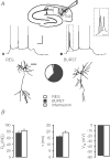
Figure 1
Figure 1. Membrane properties of subicular pyramidal cells
A, discharge behaviour upon depolarizing current pulses and reconstruction after biocytin-filling of a regular firing and a burst firing pyramidal cell, respectively. Schematic drawing illustrates placement of stimulating and recording electrodes. Recordings were conducted in the second third of the subiculum indicated by the grey shaded area. Pie chart represents 100 randomly selected neurons of which 62% were bursting (black), 35% regular firing (white) and 3% (fast-spiking) interneurons (grey). High-threshold bursting neurons and unclassified cells were excluded from the pie chart. B, neither input resistance (Rin: BURST 76 ± 6 MΩ, n = 12; REG 83 ± 6 MΩ, n = 10), nor membrane time constant (τ: BURST 16 ± 1 ms; REG 19 ± 1 ms) nor resting membrane potential (Vm BURST −57 ± 1 mV, n = 12; REG −58 ± 1 mV, n = 10) was different in regular firing and bursting neurons. Scale bars: 30 mV and 100 ms, for the inset 25 ms, and 100 μm.
Figure 2
Figure 2. LTP in regular firing cells
A, discharge pattern of a subicular regular firing cell upon negative and positive current injections. B, EPSCs recorded before and after the induction of LTP in regular firing cell shown in A. Average time course of 12 experiments. (REG – control: LTP: 1.33 ± 0.10, n = 12.) Arrow indicates tetanic stimulation.
Figure 3
Figure 3. LTP in burst firing cells
A, discharge pattern of a subicular burst firing cell upon negative and positive current injections. B, EPSCs recorded before and after the induction of LTP in burst firing cell shown in A. Pooled data for six experiments. (BURST – control: LTP: 1.59 ± 0.07, n = 6.)
Figure 4
Figure 4. LTP is blocked by the NMDA receptor antagonist APV in regular and burst firing cells
A, sharp-microelectrode recordings. Averaged time course of PTP and LTP in, A, regular firing (n = 5) and, B, bursting cells (n = 6). Superimposed traces were recorded before and after high-frequency stimulation. Scale bars: 2.5 mV and 50 ms. In both regular firing and bursting cells, LTP was blocked by the NMDA receptor antagonist APV (200 μm, REG n = 4, BURST n = 5). C, in whole-cell recordings APV also prevented LTP in bursting cells (filled circles, n = 4; open circles control, n = 6).
Figure 5
Figure 5. LTP is blocked in regular, but not in burst firing cells under voltage-clamp conditions
A, time course of EPSC amplitude in regular firing cell. Tetanus was given under voltage-clamp conditions at −60 mV. Superimposed traces were recorded before and 25–30 min after high-frequency stimulation. B, summary of six such experiments (REG – control CC: LTP: 1.33 ± 0.10, n = 12; VC: LTP: 1.04 ± 0.06, n = 6). C, time course of the EPSC amplitude in burst firing neuron. Tetanus was given while cell was voltage-clamped at −60 mV. Superimposed traces were recorded before and 25–30 min after high-frequency stimulation. D, averaged time course (BURST – control CC: LTP: 1.59 ± 0.07, n = 6; VC: LTP: 1.59 ± 0.17, n = 11). All scale bars: 100 pA and 20 ms.
Figure 6
Figure 6. Postsynaptic Ca2+ is not required for LTP in bursting cells
A, Postsynaptic dialysis with BAPTA prevented LTP in regular firing cells (0.90 ± 0.02 of baseline, n = 6). Aa shows the intrinsic firing properties and Ab the time course of an individual experiment. Scale bars Aa, 20 mV, 25 ms; Ab, 100 pA and 10 ms. B, postsynaptic Ca2+ was not required for LTP in bursting cells (1.97 ± 0.27 of baseline, n = 7). Scale bars as in A. Averaged control data for burst firing cells contain both CC and VC recordings, control data for regular firing cells only CC recordings during tetanic stimulation.
Figure 7
Figure 7. Ca2+-free conditions and BAPTA-AM block LTP in bursting cells
A, in Ca2+-free ACSF induction of LTP was blocked in bursting cells. Data for bursting cells are the same as shown in Fig. 6B, but aligned on a different time scale (0.85 ± 0.05 of baseline, n = 6). B, incubation with the cell-permeant Ca2+ chelator BAPTA-AM prevented induction of LTP in bursting neurons (0.88 ± 0.03 of baseline, n = 5).
Figure 8
Figure 8. Presynaptic expression of LTP in bursting neurons
A, EPSCs recorded in response to paired-pulse stimulation before and after tetanus-induced LTP in regular firing cells. LTP in regular firing cell (A) was not accompanied by a change in PPF (B) (1.00 ± 0.05 of baseline following high-frequency stimulation, n = 5; P = 0.99). C, EPSCs recorded in response to paired-pulse stimulation before and after tetanus-induced LTP in bursting cells. LTP in bursting cells (C) resulted in a decrease of paired-pulse facilitation (D) (0.86 ± 0.06 after tetanus, n = 9; P < 0.05).
Figure 9
Figure 9. Cell type-specific PTP at CA1-subiculum synapses
A, paired-pulse ratio at an interstimulus interval of 50 ms was not significantly different in both cell types (BURST 1.74 ± 0.21, n = 9, REG 1.53 ± 0.06, n = 5, P = 0.37). B, weak depression of EPSCs in both cell types was observed using a stimulus protocol of 100 pulses at 50 Hz. Ca, cell type-specific PTP induced by 100 pulses at 50 Hz in regular and burst firing cells in the absence of LTP. Representative examples. Scale bars: 200 pA and 10 ms. Cb, averaged PTP recorded 5 s after the stimulus was significantly stronger in bursting cells than in regular firing cells (BURST 3.08 ± 0.27, n = 9; REG 1.79 ± 0.09, n = 9; P < 0.01). Open arrows indicate repetitive stimulation of 100 pulses at 50 Hz.

References

    1. Amaral DG, Witter MP. The hippocampal formation. In: Paxinos G, editor. The Rat Nervous System. New York: Academic Press; 1995. pp. 443–494.
    1. Bliss TV, Collingridge GL. A synaptic model of memory: long-term potentiation in the hippocampus. Nature. 1993;361:31–39. - PubMed
    1. Boeijinga PH, Boddeke HW. Activation of 5-HT1B receptors suppresses low but not high frequency synaptic transmission in the rat subicular cortex in vitro. Brain Res. 1996;721:59–65. - PubMed
    1. Brasier DJ, Feldman DE. Synapse-specific expression of functional presynaptic NMDA receptors in the rat somatosensory cortex. J Neurosci. 2008;28:2199–2211. - PMC - PubMed
    1. Casado M, Isope P, Ascher P. Involvement of presynaptic N-methyl-D-aspartate receptors in cerebellar long-term depression. Neuron. 2002;33:123–130. - PubMed

Publication types

LinkOut - more resources