Clinical Trial
. 2008 Jun;10(3):320-9.
doi: 10.1215/15228517-2008-012. Epub 2008 Apr 10.

Intracerebral infusion of an EGFR-targeted toxin in recurrent malignant brain tumors

Affiliations
Clinical Trial

Intracerebral infusion of an EGFR-targeted toxin in recurrent malignant brain tumors

John H Sampson et al. Neuro Oncol. 2008 Jun.

Abstract

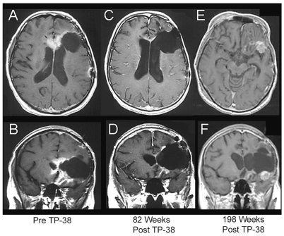
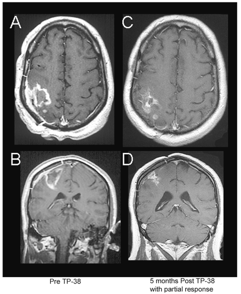
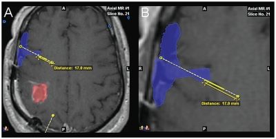
The purpose of this study is to determine the maximum tolerated dose (MTD), dose-limiting toxicity (DLT), and intracerebral distribution of a recombinant toxin (TP-38) targeting the epidermal growth factor receptor in patients with recurrent malignant brain tumors using the intracerebral infusion technique of convection-enhanced delivery (CED). Twenty patients were enrolled and stratified for dose escalation by the presence of residual tumor from 25 to 100 ng/ml in a 40-ml infusion volume. In the last eight patients, coinfusion of (123)I-albumin was performed to monitor distribution within the brain. The MTD was not reached in this study. Dose escalation was stopped at 100 ng/ml due to inconsistent drug delivery as evidenced by imaging the coinfused (123)I-albumin. Two DLTs were seen, and both were neurologic. Median survival after TP-38 was 28 weeks (95% confidence interval, 26.5-102.8). Of 15 patients treated with residual disease, two (13.3%) demonstrated radiographic responses, including one patient with glioblastoma multiforme who had a nearly complete response and remains alive >260 weeks after therapy. Coinfusion of (123)I-albumin demonstrated that high concentrations of the infusate could be delivered >4 cm from the catheter tip. However, only 3 of 16 (19%) catheters produced intraparenchymal infusate distribution, while the majority leaked infusate into the cerebrospinal fluid spaces. Intracerebral CED of TP-38 was well tolerated and produced some durable radiographic responses at doses <or=100 ng/ml. CED has significant potential for enhancing delivery of therapeutic macromolecules throughout the human brain. However, the potential efficacy of drugs delivered by this technique may be severely constrained by ineffective infusion in many patients.


Figures

Fig. 1
Fig. 1
Axial and coronal MR images of a 59-year-old female with a recurrent glioblastoma multiforme (GBM) (patient 2) showing a sustained radiographic response to TP-38. This patient had failed surgery, external beam radiation therapy, carmustine, cisplatin, etoposide, temozolomide with carmustine wafers, irinotecan, cyclophosphamide, and tamoxifen prior to receiving TP-38 therapy. Serial images from left to right show her scans before TP-38 therapy (A, B); 82 weeks after TP-38 with a nearly complete response (C, D); and, most recently, 198 weeks after TP-38 with biopsy-proven, recurrent GBM (E, F). She remains working full time with a KPS of 90.
Fig. 2
Fig. 2
Axial and coronal MR images of a 48-year-old male with recurrent glioblastoma multiforme (patient 18) showing a partial radiographic response to TP-38. This patient had failed prior surgery and external beam radiation therapy. Images show his scans before TP-38 therapy (A, B) and 5 months after therapy with a partial response (C, D). He subsequently died from an unrelated sepsis. Up to that time, however, he was working full time with a KPS of 90.
Fig. 3
Fig. 3
Axial and coronal MR images of a 55-year-old female with recurrent glioblastoma multiforme (patient 12) showing delayed enhancement. Serial images from left to right show her scans postoperatively and before TP-38 therapy (A, B); 6 weeks after therapy (C, D); 11 months after therapy with a new area of contrast enhancement (E, F); 24 months after therapy with partial spontaneous resolution of this area of enhancement without treatment (G, H); 30 months after therapy with further resolution of the contrast-enhancing area (I, J); and most recently, 43 months after TP-38 therapy with nearly complete resolution of all enhancing areas (K, L).
Fig. 4
Fig. 4
Co-registered images showing optimal distribution of 123I-albumin after convection-enhanced delivery. (A) Single-photon emission computed tomography (SPECT) image of 123I-albumin distribution co-registered with axial MR image for anterior right frontal catheter positioned below the resection cavity (patient 12). (B) SPECT image of 123I-albumin distribution co-registered with axial MR image for posterior right frontal catheter positioned behind the resection cavity. (C, D) Corresponding SPECT-derived isodose contours co-registered with axial T1-weighted MR images located at the tip of each catheter in the right frontal lobe of the brain. Isodose lines show the percent concentration of 123I-albumin relative to the infused concentration (0.25 mg/ml) such that at the 50% isodose line the concentration of albumin is 0.125 mg/ml and at the 10% isodose line the concentration of albumin is 0.025 mg/ml. (E, F) Oblique reconstructions of complete SPECT image showing areas covered and not covered by the infusion.
Fig. 5
Fig. 5
Co-registered images showing ineffective distribution of 123I-albumin after convection-enhanced delivery: selected axial MR image (A) and coronal images (B, C) from three different patients showing single-photon emission computed tomography image of 123I-albumin distribution co-registered with MR images demonstrating leakage of infusate into the subarachnoid cerebrospinal fluid spaces over the superior hemispheric surface of frontoparietal lobe of the brain (A–C) and along the tentorial surface of the occipital lobe (C).
Fig. 6
Fig. 6
Co-registered images showing distribution of 123I-albumin into the Sylvian fissure after convection-enhanced delivery from a catheter with proximal ports: outline (A) and magnified view (B) of single-photon emission computed tomography image of 123I-albumin distribution (blue) shown co-registered with axial MR image (patient 18). The tumor is outlined in red. A second catheter trajectory is shown in yellow at the bottom of A. The most proximal port on this catheter is located 17.0 mm from the tip. In this instance, the infusate appears to have exited the catheter through the most proximal port, which was communicating with the subarachnoid cerebrospinal fluid (CSF) space within the deep portion of the Sylvian fissure between the frontal and temporal lobes of the brain. This allowed the infusate to leak completely into the CSF spaces without any penetration of the parenchyma.

Similar articles

Cited by

References

    1. CBTRUS, Central Brain Tumor Registry of the United States. Statistical Report: Primary Brain Tumors in the United States, 1997–2001. Chicago: CBTRUS; 2004.
    1. Imperato JP, Paleologos NA, Vick NA. Effects of treatment on long-term survivors with malignant astrocytomas. Ann Neurol. 1990;28:818–822. - PubMed
    1. Pai LH, Pastan I. Immunotoxins and recombinant toxins. In: DeVita VT Jr, Hellman S, Rosenberg SA, editors. Biologic Therapy of Cancer. Philadelphia: J.B. Lippincott; 1995. pp. 521–533.
    1. Phillips PC, Levow C, Catterall M, et al. Transforming growth factor-alpha-Pseudomonas exotoxin fusion protein (TGF-alpha-PE38) treatment of subcutaneous and intracranial human glioma and medulloblastoma xenografts in athymic mice. Cancer Res. 1994;54:1008–1015. - PubMed
    1. Pastan I. Targeted therapy of cancer with recombinant immunotoxins. Biochimica et Biophysica Acta. 1997;1333:C1–C6. - PubMed

Publication types

MeSH terms

Substances