. 2008 Apr 11;320(5873):239-43.
doi: 10.1126/science.1155313.

Leiomodin is an actin filament nucleator in muscle cells

Affiliations

Leiomodin is an actin filament nucleator in muscle cells

David Chereau et al. Science. .

Abstract

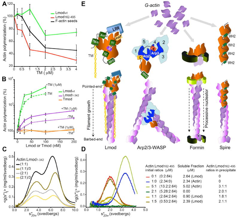
Initiation of actin polymerization in cells requires nucleation factors. Here we describe an actin-binding protein, leiomodin, that acted as a strong filament nucleator in muscle cells. Leiomodin shared two actin-binding sites with the filament pointed end-capping protein tropomodulin: a flexible N-terminal region and a leucine-rich repeat domain. Leiomodin also contained a C-terminal extension of 150 residues. The smallest fragment with strong nucleation activity included the leucine-rich repeat and C-terminal extension. The N-terminal region enhanced the nucleation activity threefold and recruited tropomyosin, which weakly stimulated nucleation and mediated localization of leiomodin to the middle of muscle sarcomeres. Knocking down leiomodin severely compromised sarcomere assembly in cultured muscle cells, which suggests a role for leiomodin in the nucleation of tropomyosin-decorated filaments in muscles.


Figures

Fig. 1
Fig. 1
Domain organization and localization of Lmod in cultured rat cardiomyocytes. (A) Modular organization of Lmod and constructs used in this study (TM-h1, TM-h2 and A-h, tropomyosin and actin binding helices; polyP, poly-proline; h1 and h2, predicted helices; B, basic segment). (B) Fluorescent antibody staining shows colocalization of endogenous Lmod (green) and myomesin (red) in the middle of sarcomeres. (C) EGFP-Lmodwt and EGFP-Lmod1-342 constructs (green) also concentrate in the middle of sarcomeres, separated from Z-discs visualized with antibodies to α-actinin, whereas EGFP-Lmod162-495 concentrates in the nucleus (bar, 10μm).
Fig. 2
Fig. 2
Lmod knockdown disrupts sarcomere assembly in rat cardiomyocytes. Lmod antibody staining is reduced and the striated Lmod pattern is lost in cardiomyocytes transfected with Lmod specific siRNA as compared to control cells (transfected with scrambled-siRNA). The micrographs of Alexa 568-phalloidin show that sarcomeric actin structures are severely disorganized in knockdown cells (bar, 10μm).
Fig. 3
Fig. 3
Lmod nucleates actin filaments. (A) Time course of fluorescence increase upon polymerization of 2μM Mg-ATP-actin (6% pyrene-labeled) alone (black) or with 25nM Lmod constructs (colored according to Fig. 1A) in polymerization buffer (10mM Tris pH7.5, 1mM MgCl2, 50mM KCl, 1mM EGTA, 0.1mM NaN3, 0.02mg/mL BSA, 0.2mM ATP). (B) Number of barbed-ends formed after 4min of polymerization with varying Lmod concentration measured by TIRF. For each construct, the average number of actin seeds was measured from 20 images (150 × 120 μm). Data reported are mean ± SD. (C) TIRF micrographs of fields of actin filaments prepared 4min after initiating of polymerization of 1μM Mg-ATP-actin with a range of Lmodwt concentrations. (D) Time course of barbed end formation in the presence of Lmod constructs calculated from the polymerization rate, concentration of actin monomers remaining, and rate constant for barbed-end elongation (10 μM-1s-1) (fig. S7A). The table gives the nucleation rates.
Fig. 4
Fig. 4
Mechanism of actin nucleation by Lmod. (A) Effect of tropomyosin on the polymerization of 2μM actin induced by 0.5μM F-actin seeds, 25nM Lmodwt or 25nM Lmod162-495. Rates were determined from the slope of the curves at 50% polymerization and expressed as percentage of the rates without tropomyosin. Data reported are mean ± SD of three or more experiments. (B) Polymerization rates of 2μM actin with increasing concentrations of Lmodwt, Lmod1-342 or Tmod in the absence (dashed lines) or the presence (solid lines) of 1μM tropomyosin. Rates are expressed as percentages of the polymerization rate of actin alone. Data reported are mean ± SD of three or more experiments. (C) Sedimentation velocity dilution series of complexes of actin:Lmod1-342 at two different molar ratios. The samples contained 50μM LatB to prevent polymerization. The first peak corresponds to actin-LatB. The main species in the second peak is a 2:1 actin:Lmod1-342 complex. (D) Sedimentation velocity analysis of actin:Lmod162-495 mixed at different ratios (right). In the presence of LatB, actin:Lmod162-495 complexes precipitated. The stoichiometries of the complexes in the precipitate were determined by difference from the amount of actin or Lmod162-495 remaining in solution, using the interference optics of the ultracentrifuge. (E) Comparison of models for actin filament nucleation. We propose that Lmod stabilizes a trimeric actin nucleus and that tropomyosin enhances nucleation and mediates the localization of Lmod to filament pointed-ends. Arp2 and Arp3 of Arp2/3 complex form a trimeric seed with an actin monomer bound to the WH2 of a nucleation-promoting factor. Formin dimers form nuclei and remain associated with the barbed-end of the growing filament. Spire stabilizes a nucleus consisting of four actin subunits along a long-pitch filament strand.

Similar articles

Cited by

References

    1. Pollard TD. Annu Rev Biophys Biomol Struct. 2007;36:451. - PubMed
    1. Fischer RS, Fowler VM. Trends Cell Biol. 2003;13:593. - PubMed
    1. Conley CA, Fritz-Six KL, Almenar-Queralt A, Fowler VM. Genomics. 2001;73:127. - PubMed
    1. Kostyukova AS, Hitchcock-Degregori SE, Greenfield NJ. J Mol Biol. 2007;372:608. - PMC - PubMed
    1. Fowler VM, Greenfield NJ, Moyer J. J Biol Chem. 2003;278:40000. - PubMed

Publication types

MeSH terms