. 2008 May 23;102(10):1247-55.
doi: 10.1161/CIRCRESAHA.108.172379. Epub 2008 Apr 10.

Modulation of endothelial cell KCa3.1 channels during endothelium-derived hyperpolarizing factor signaling in mesenteric resistance arteries

Affiliations

Modulation of endothelial cell KCa3.1 channels during endothelium-derived hyperpolarizing factor signaling in mesenteric resistance arteries

Kim A Dora et al. Circ Res. .

Abstract

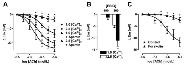
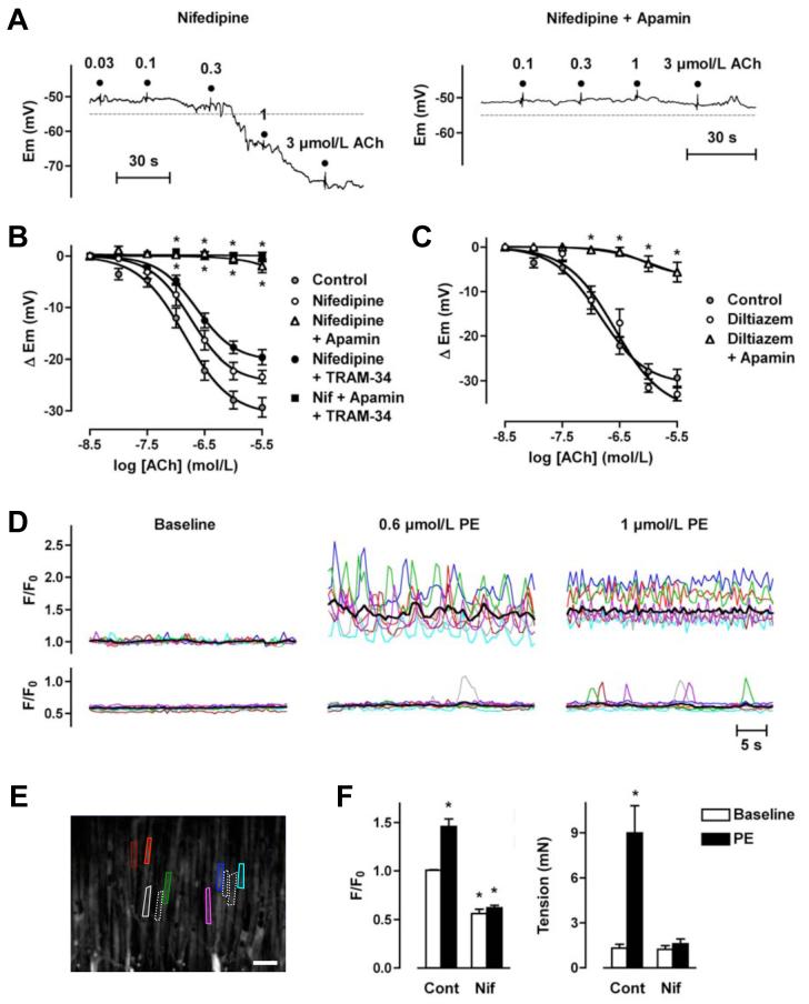
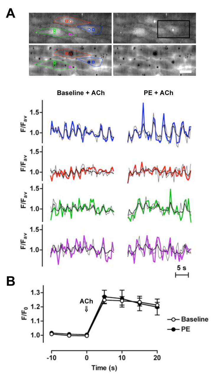
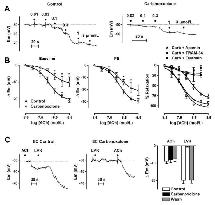
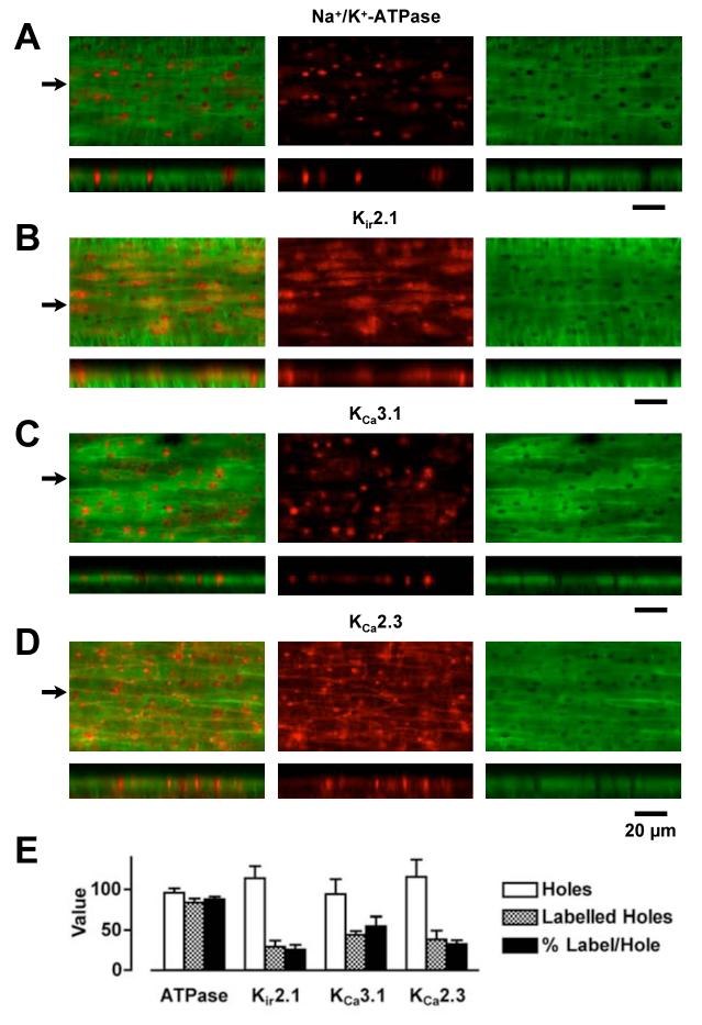
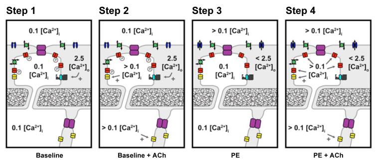
Arterial hyperpolarization to acetylcholine (ACh) reflects coactivation of K(Ca)3.1 (IK(Ca)) channels and K(Ca)2.3 (SK(Ca)) channels in the endothelium that transfers through myoendothelial gap junctions and diffusible factor(s) to affect smooth muscle relaxation (endothelium-derived hyperpolarizing factor [EDHF] response). However, ACh can differentially activate K(Ca)3.1 and K(Ca)2.3 channels, and we investigated the mechanisms responsible in rat mesenteric arteries. K(Ca)3.1 channel input to EDHF hyperpolarization was enhanced by reducing external [Ca(2+)](o) but blocked either with forskolin to activate protein kinase A or by limiting smooth muscle [Ca(2+)](i) increases stimulated by phenylephrine depolarization. Imaging [Ca(2+)](i) within the endothelial cell projections forming myoendothelial gap junctions revealed increases in cytoplasmic [Ca(2+)](i) during endothelial stimulation with ACh that were unaffected by simultaneous increases in muscle [Ca(2+)](i) evoked by phenylephrine. If gap junctions were uncoupled, K(Ca)3.1 channels became the predominant input to EDHF hyperpolarization, and relaxation was inhibited with ouabain, implicating a crucial link through Na(+)/K(+)-ATPase. There was no evidence for an equivalent link through K(Ca)2.3 channels nor between these channels and the putative EDHF pathway involving natriuretic peptide receptor-C. Reconstruction of confocal z-stack images from pressurized arteries revealed K(Ca)2.3 immunostain at endothelial cell borders, including endothelial cell projections, whereas K(Ca)3.1 channels and Na(+)/K(+)-ATPase alpha(2)/alpha(3) subunits were highly concentrated in endothelial cell projections and adjacent to myoendothelial gap junctions. Thus, extracellular [Ca(2+)](o) appears to modify K(Ca)3.1 channel activity through a protein kinase A-dependent mechanism independent of changes in endothelial [Ca(2+)](i). The resulting hyperpolarization links to arterial relaxation largely through Na(+)/K(+)-ATPase, possibly reflecting K(+) acting as an EDHF. In contrast, K(Ca)2.3 hyperpolarization appears mainly to affect relaxation through myoendothelial gap junctions. Overall, these data suggest that K(+) and myoendothelial coupling evoke EDHF-mediated relaxation through distinct, definable pathways.


Figures

Figure 1
Figure 1
Summarized data showing the average change in membrane potential (Δ Em) to cumulative increases in [ACh] applied to evoke EDHF-hyperpolarization in mesenteric arteries. A, Reducing [Ca2+]o from 2.5 mmol/L to 1.0 mmol/L depressed the steady state hyperpolarization to ACh (n=7 & 12, respectively). In 1.0 mmol/L Ca2+ and with 50 nmol/L apamin present approximately 50% hyperpolarization remained (n=4), whereas in 2.5 mmol/L [Ca2+]o, apamin fully blocked hyperpolarization (n=13). B, Histogram summarizing the increase in smooth muscle membrane potential following endothelial cell stimulation with 1-EBIO in the presence of 50 nmol/L apamin. In 1.0 mmol/L [Ca2+]o, hyperpolarization to both 100 and 300 μmol/L 1-EBIO was enhanced (n=4&5, respectively). Asterisk indicates significant difference relative to control (2.5 mmol/L [Ca2+]o, P<0.05). C, In 1.0 mmol/L [Ca2+]o Krebs with 50 nmol/L apamin present EDHF-hyperpolarization evoked with ACh was significantly inhibited in the additional presence of 1 μmol/L forskolin (n=8, P<0.05).
Figure 2
Figure 2
ACh-mediated stimulation of KCa3.1-channels is prevented by blocking VGCC in rat mesenteric arteries. A, Original traces demonstrating concentration-dependent EDHF-induced smooth muscle hyperpolarization evoked by ACh in the presence of nifedipine (10 μmol/L) and prior depolarization to PE. Hyperpolarization overshot the resting membrane potential (dashed line), and was abolished by apamin (50 nmol/L). B, Summarized data showing the average change in membrane potential (Δ Em) to cumulative increases in [ACh] in arteries stimulated with PE in the presence of nifedipine (10 μmol/L, n=4-10), or in the additional presence of either apamin (50 nmol/L, n=4-12) or TRAM-34 (1 μmol/L, n=7-14), or both inhibitors together (n=5-8). C, Summarized data showing the average change in membrane potential (Δ Em) to cumulative increases in [ACh] in arteries stimulated with PE in the presence of diltiazem (10 μmol/L, n=4-6), or in the additional presence of apamin (50 nmol/L, n=5-7). D, Smooth muscle Ca2+ signals in the absence (upper traces) and presence of nifedipine (10 μmol/L, lower traces) colour-coded to the field of interest shown in the image in E. Applying 0.6 or 1 μmol/L PE stimulated a marked increase in the global average level of [Ca2+] (black line), representing asynchronous Ca2+ increases within individual muscle cells. Both the average increase and the majority of the oscillations were abolished in the presence of nifedipine (see Online Video 1 for movie corresponding to these data). F, Summarized data showing inhibition of the increase in [Ca2+]i stimulated by 1 μmol/L PE (left panel, control n=6, nifedipine n=3) and the associated block of arterial contraction (right panel, paired data). Asterisk indicates significant difference relative to control baseline (P<0.05).
Figure 3
Figure 3
Similar increases in endothelial cell [Ca2+]i evoked with ACh alone and applied during smooth muscle stimulation with PE. A, Mesenteric arteries were mounted in a pressure myograph and changes in endothelial cell [Ca2+]i assessed at the interface between the endothelium and the IEL. Upper micrographs show loaded endothelial cells (average of 20 sec). Note bright spots (endothelial cell projections) that correspond to holes in the IEL (lower micrographs). Bar = 20 μm. Lower panels show the time course of fluorescence changes in individual endothelial cells in response to ACh (1 μmol/L) added under baseline conditions (left) and in the presence of PE (0. 3 - 0.6 μmol/L, right). Regions were placed over (colour) and adjacent to (grey) the endothelial cell projections, and around the whole cell (black). The colours relate to subcellular regions within the micrographs (see Online Video 2 for movie corresponding to these data from the region indicated by the black box). Representative of 3 experiments. B, Mesenteric arteries were mounted in a wire myograph and changes in (whole) endothelial cell [Ca2+]i measured before and during ACh (1 μmol/L) either from the baseline or in the presence of PE (0.3 - 0.6 μM, n=3, paired data). Note that endothelial cell projections were not loaded in these experiments (see Online Methods Supplement for details).
Figure 4
Figure 4
Gap junctions facilitate the contribution of KCa2.3-channels to EDHF-hyperpolarization in rat mesenteric artery. A, Original traces demonstrating ACh-mediated concentration-dependent increases in smooth muscle cell membrane potential from the resting membrane potential (dashed lines). Carbenoxolone (100 μmol/L) did not alter the resting membrane potential, but reduced the hyperpolarization to ACh. B, Summarized data showing the average change in membrane potential (Δ Em) to cumulative increases in [ACh] to evoke EDHF-hyperpolarization in arteries from resting membrane potential (left, n=6-11). In the presence of PE (right panels), carbenoxolone (100 μmol/L) depressed EDHF hyperpolarization (n=5-9) but not relaxation to ACh (n=8-14). The EDHF-mediated relaxation was reduced by the addition of apamin (50 nmol/L, n=9-13), and almost abolished by TRAM-34 (1 μmol/L, n=5-12) or ouabain (100 μmol/L, n=3-9), each applied when carbenoxolone was present. Asterisk indicates significant difference relative to control (P<0.05).
Figure 5
Figure 5
Endothelial cell immunohistochemical expression pattern for Na+/K+-ATPase, Kir2.1-, KCa3.1- and KCa2.3-channels in rat mesenteric arteries. (A-D, top panels) Confocal images of the wall of pressurized arteries showing a single z-axis plane through the IEL (green) and simultaneously acquired corresponding expression of protein (red). (A-D, bottom panels) Reconstruction of the confocal z-axis multi-plane stack through the wall of the artery corresponding to a line drawn through the images in the upper panels at the positions indicated by arrows. Note the presence of protein staining within the holes through the IEL in both the upper and lower panels. Both the Na+/K+-ATPase α2-subunit and KCa3.1-channels are highly expressed within the holes, whereas Kir2.1- and KCa2.3-channels are also highly expressed within endothelial cells and at endothelial cell borders, respectively. In these images, the Alexa Fluor 488 secondary antibody was only applied to the luminal side of the artery, and the confocal laser and PMT settings were identical for each z-stack. Bar indicates 20 μm in the x-axis in all images. Representative of at least 3 arteries. E, Quantification of protein expression within the holes through the IEL (n=3-5).
Figure 6
Figure 6
Suggested key steps underlying endothelial cell KCa-channel activation and subsequent transfer of hyperpolarization to the mesenteric smooth muscle. Step 1. At rest in 2.5 mmol/L [Ca2+]o Krebs solution, the endothelial cell Ca2+ sensing receptor (CaSR) is maximally stimulated and KCa3.1-channels are in some way inactivated, possibly via PKA-phosphorylation. Step 2. Stimulation of the endothelium with ACh raises cytoplasmic [Ca2+]i, activating apamin-sensitive KCa2.1-channels causing hyperpolarization which can spread through homocellular (endothelial-endothelial) and heterocellular (myoendothelial, MEGJs) gap junctions. KCa3.1 are not available for agonist-induced [Ca2+]i activation. Step 3. Activation of smooth muscle VDCC (with PE) causes a local ‘sink’ of [Ca2+]o in the vicinity of the endothelial projections. This reduction reduces stimulation of the CaSR and phosphorylation of KCa3.1-channels in the projections. Step 4. At this point, ACh-stimulation of the endothelium raises cytoplasmic [Ca2+]i, now activating ‘available’ KCa3.1 as well as KCa2.3. As in Step 2, the resulting hyperpolarization spreads through endothelial-endothelial and MEGJs gap junctions, but now reflects input from both KCa2.3 and KCa3.1. Facilitated by ongoing muscle depolarization, endothelial K+ efflux through KCa-channels is sufficient to stimulate adjacent Na+/K+-ATPase enhancing hyperpolarization. Further amplification may also occur through KIR-channel activation within this microdomain. As smooth muscle cells repolarize to resting levels, VGCC open-probability decreases, removing the Ca2+ ‘sink’, local [Ca2+]o increases towards 2.5 mmol/L and KCa3.1-channel activity is again removed from the control of intracellular [Ca2+]i. Grey, intercellular space; Purple, gap junctions; red, KCa3.1-channels; yellow, KCa2.3-channels; blue, VGCCs; green, Na+/K+-ATPase; and cyan, CaSR.

Comment in

  • The EDHF story: the plot thickens.
    Parkington HC, Tare M, Coleman HA. Parkington HC, et al. Circ Res. 2008 May 23;102(10):1148-50. doi: 10.1161/CIRCRESAHA.108.177279. Circ Res. 2008. PMID: 18497313 No abstract available.

Similar articles

Cited by

References

    1. Busse R, Edwards G, Feletou M, Fleming I, Vanhoutte PM, Weston AH. EDHF: bringing the concepts together. Trends Pharmacol Sci. 2002;23:374–380. - PubMed
    1. Burnham MP, Johnson IT, Weston AH. Impaired small-conductance Ca2+-activated K+ channel-dependent EDHF responses in Type II diabetic ZDF rats. Br J Pharmacol. 2006;148:434–441. - PMC - PubMed
    1. Kohler R, Wulff H, Eichler I, Kneifel M, Neumann D, Knorr A, Grgic I, Kampfe D, Si H, Wibawa J, Real R, Borner K, Brakemeier S, Orzechowski HD, Reusch HP, Paul M, Chandy KG, Hoyer J. Blockade of the intermediate-conductance calcium-activated potassium channel as a new therapeutic strategy for restenosis. Circulation. 2003;108:1119–1125. - PubMed
    1. Crane GJ, Gallagher NT, Dora KA, Garland CJ. Small and intermediate calcium-dependent K+ channels provide different facets of endothelium-dependent hyperpolarization in rat mesenteric artery. J Physiol. 2003;553:183–189. - PMC - PubMed
    1. Sandow SL, Neylon CB, Chen MX, Garland CJ. Spatial separation of endothelial small- and intermediate-conductance calcium-activated potassium channels (K(Ca)) and connexins: possible relationship to vasodilator function? J Anat. 2006;209:689–698. - PMC - PubMed

Publication types

MeSH terms