. 2009 Jan;27(1):116-25.
doi: 10.1634/stemcells.2007-0205.

Efficient serum-free derivation of oligodendrocyte precursors from neural stem cell-enriched cultures

Affiliations

Efficient serum-free derivation of oligodendrocyte precursors from neural stem cell-enriched cultures

Rajesh C Rao et al. Stem Cells. 2009 Jan.

Abstract

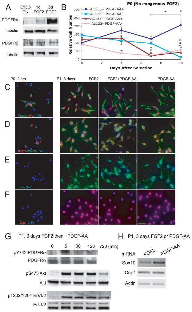
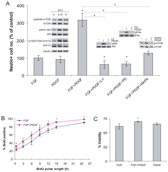
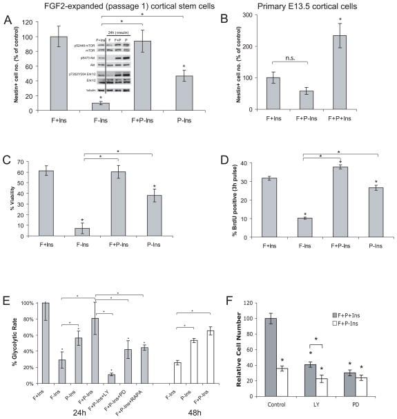
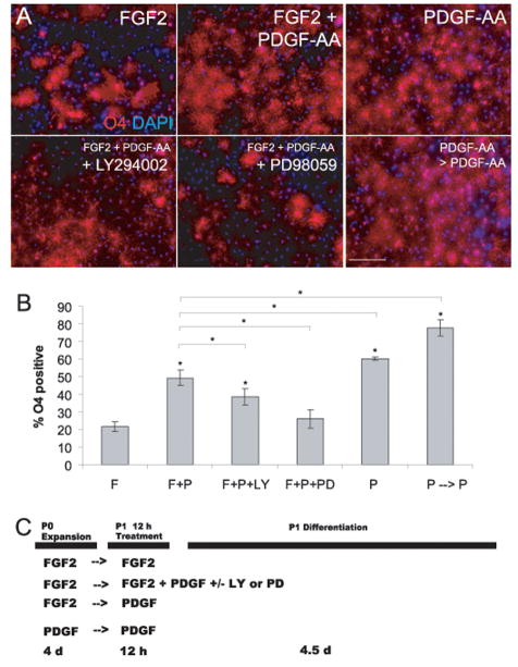
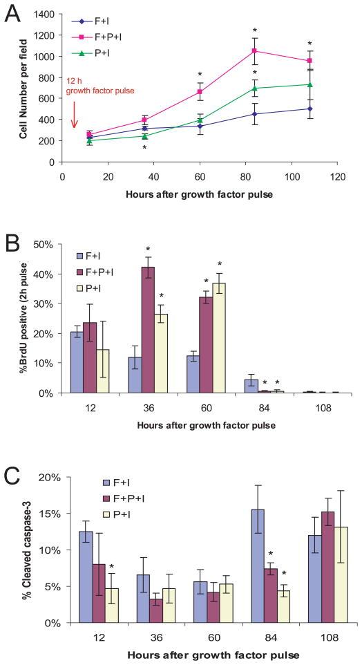
Oligodendrocytes derived in the laboratory from stem cells have been proposed as a treatment for acute and chronic injury to the central nervous system. Platelet-derived growth factor (PDGF) receptor alpha (PDGFRalpha) signaling is known to regulate oligodendrocyte precursor cell numbers both during development and adulthood. Here, we analyze the effects of PDGFRalpha signaling on central nervous system (CNS) stem cell-enriched cultures. We find that AC133 selection for CNS progenitors acutely isolated from the fetal cortex enriches for PDGF-AA-responsive cells. PDGF-AA treatment of fibroblast growth factor 2-expanded CNS stem cell-enriched cultures increases nestin(+) cell number, viability, proliferation, and glycolytic rate. We show that a brief exposure to PDGF-AA rapidly and efficiently permits the derivation of O4(+) oligodendrocyte-lineage cells from CNS stem cell-enriched cultures. The derivation of oligodendrocyte-lineage cells demonstrated here may support the effective use of stem cells in understanding fate choice mechanisms and the development of new therapies targeting this cell type.


Conflict of interest statement

Disclosures

The authors indicate no potential conflicts of interest.

Figures

Figure 1
Figure 1. Fetal CNS stem cell-enriched cultures are PDGF-AA responsive: PDGF-AA treatment activates PI3K/Akt and MEK/Erk pathways but maintains expression of precursor markers
(A) Western blot (WB) analysis of PDGFRs α and β in E13.5 lateral cortex, and after 3 and 5 day treatment with FGF2. (B) AC133 selection enriches for PDGF-AA responsive cells. Acutely dissected E13.5 cortical cells were sorted based on expression of AC133, and positively and negatively selected cells were cultured (plated at 12,500 cells/cm2) with or without PDGF-AA (no exogenous FGF2 was added) and cell number (mean ± s.e.m., n = 5 per timepoint, *p < 0.05) was measured at 0 (30 min after plating), 4, 7, and 10 d. (C,D,E,F) Passage 1, FGF2-expanded, CNS stem cells with treated with FGF2 alone, FGF2 + PDGF-AA, or switched from FGF2 to PDGF-AA alone for 3 d. Primary (Passage 0) E13.5 cortical cells, untreated with FGF2 or PDGF-AA, serve as controls. PDGF-AA treated cells maintained expression of several precursor markers, including (C) nestin (green), Sox2 (red); (D) A2B5 (red), PDGFRα (green); (E) NG2 (green); (F) Olig2 (red). All nuclei (B–F) were stained with DAPI (blue). Scale bar = 20 μm. (G) PDGF-AA causes phosphorylation of PDGFRα and rapid and sustained activation of MEK/Erk and PI3K/Akt pathways. (H) RT-PCR data for Sox10, Cnp1, and Actin mRNA after 3 d treatment of passage 1 CNS stem cells with FGF2 or PDGF-AA.
Figure 2
Figure 2. Co-stimulation with PDGF-AA increases nestin+ cell number, viability, and proliferation; and activates mTOR PI3K/Akt, MEK/Erk pathways
(A) CNS stem cell-enriched cultures were plated at low density (5,000 cells/cm2) and grown for 4 d ± FGF2, PDGF-AA, inhibitors of PI3K (LY, LY294002, 10 μM), MEK (PD, PD98059, 50 μM) and mTOR (RAPA, rapamycin, 1μM). Cell number (mean ± s.d., n = 4, *p < 0.03) is expressed as percent of control (FGF2). WB analyses confirm inhibition of mTOR PI3K/Akt, MEK/Erk pathways. (B) CNS stem cell-enriched cultures treated with FGF2 alone vs. FGF2 and PDGF-AA were pulsed with 10 μM BrdU for increasing time periods, fixed, and labeled with anti-BrdU antibody (mean ± s.d., n = 3, *p < 0.015). To compare S-phase entry of control cells with cells switched from FGF2 to PDGF-AA, cells were pulsed for 24 h, and labeled with anti-BrdU antibody (mean ± s.d., n = 3 per timepoint, n.s. = non-significant). (C) CNS stem cell-enriched cultures were grown for 3 d ± FGF2, PDGF-AA, and viability was analyzed based on propidium-iodide exclusion using FACS (n = 3, *p < 0.05).
Figure 3
Figure 3. PDGF-AA rescues nestin+ cell number, viability, and proliferation in the absence of FGF2 and insulin and increases glycolytic rate
(A) CNS stem cell-enriched cultures were plated at low density and grown for 4 d ± FGF2 (F), PDGF-AA (P), and insulin (Ins). Cell number (mean ± s.d., n = 4, *p < 0.001) is expressed as percent of control (FGF2). (B) Freshly dissected E13.5 primary cortical cells were plated at low density and grown for 4 d ± FGF2, PDGF-AA, and insulin (mean ± s.e.m., n.s. = non-significant [p = 0.09], *p < 0.02). (C) CNS stem cell-enriched cultures were grown for 3 d ± FGF2, PDGF-AA, and insulin; viability was analyzed based on propidium-iodide exclusion using FACS (mean ± s.d., n = 3, *p < 0.003). (D) CNS stem cell-enriched cultures were pulsed with BrdU for 3 h, fixed and then labeled with an anti-BrdU antibody (mean ± s.d., n = 3, *p < 0.003). (E) CNS stem cell-enriched cultures were cultured for 24 or 48 h ± F, P, Ins, LY, PD, or RAPA and glycolytic rate was measured. Glycolytic rate (mean ± s.d., n = 4, *p < 0.015) is expressed as percent of control (F+Ins). (F) CNS stem cell-enriched cultures were plated at low density and grown for 4 d with FGF2 and PDGF-AA and ± insulin and inhibitors of PI3K (LY, LY294002, 10 μM), MEK (PD, PD98059, 50 μM) (mean ± s.d., n = 3).
Figure 4
Figure 4. PDGF-AA promotes oligodendrogliogenesis from CNS stem cell-enriched cultures
(A) CNS stem cells were plated at 25,000 cells/cm2 and pulsed for 12 h ± FGF2, PDGF-AA, LY, and/or PD (cultures were pretreated with inhibitors for 1 h prior to PDGF-AA pulse). Cells in “PDGF-AA only” group were obtained by plating primary E13.5 cortical cells in PDGF-AA alone (no exogeneous FGF2 was added) for 5–7 d, passaging, and plating at same density as FGF2-expanded CNS stem cells. This enriched population of oligodendrocyte precursors was treated with PDGF-AA for an additional 12 h. After cytokine and/or inhibitor treatment, cells were then switched to serum-free differentiation medium, cultured for 4–5 d, fixed, and labeled with anti-O4 antibody. Scale bar = 20 μm. (B) Percentage of O4+ oligodendrocytes of total cells (mean ± s.d., n = 4 [n = 3 for inhibitor-treated groups], *p < 0.025). (C) Treatment scheme used in (A) and (B)
Figure 5
Figure 5. Transient exposure to PDGF-AA is associated with a delay in cell cycle exit during differentiation
(A) CNS stem cells were treated as in Fig 4A. Twelve, 36, 60, 84, and 108 hours after growth factor pulse, differentiating cells were fixed (BrdU was added 2 h before fixation as indicated), and cell number, (B) S-phase, and (C) apoptosis was measured by labeling with DAPI, anti-BrdU, and anti-cleaved caspase-3 antibodies (mean ± s.d., n = 3 for each timepoint, *p < 0.04).

Similar articles

Cited by

References

    1. McKay R. Stem cells in the central nervous system. Science. 1997 Apr 4;276(5309):66–71. - PubMed
    1. Gage FH. Mammalian neural stem cells. Science. 2000 Feb 25;287(5457):1433–1438. - PubMed
    1. Uchida N, Buck DW, He D, et al. Direct isolation of human central nervous system stem cells. Proc Natl Acad Sci U S A. 2000 Dec 19;97(26):14720–14725. - PMC - PubMed
    1. Okabe S, Forsberg-Nilsson K, Spiro AC, Segal M, McKay RD. Development of neuronal precursor cells and functional postmitotic neurons from embryonic stem cells in vitro. Mech Dev. 1996 Sep;59(1):89–102. - PubMed
    1. Zhang SC, Wernig M, Duncan ID, Brustle O, Thomson JA. In vitro differentiation of transplantable neural precursors from human embryonic stem cells. Nat Biotechnol. 2001 Dec;19(12):1129–1133. - PubMed

Publication types

MeSH terms