

Different mechanisms of DEHP-induced hepatocellular adenoma tumorigenesis in wild-type and Ppar alpha-null mice
- PMID: 18403868
- PMCID: PMC7217336
- DOI: 10.1539/joh.l7105
Different mechanisms of DEHP-induced hepatocellular adenoma tumorigenesis in wild-type and Ppar alpha-null mice
Abstract
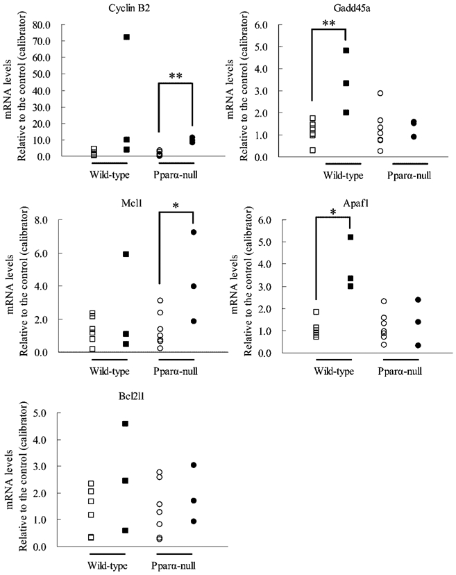
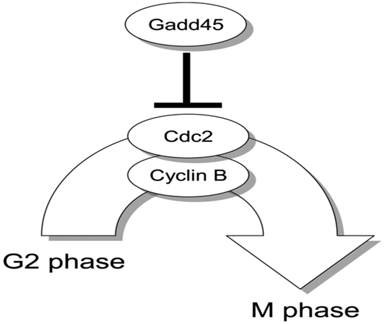
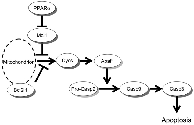
Di (2-ethylhexyl) phthalate (DEHP) exposure is thought to lead to hepatocellular hypertrophy and hyperplasia in rodents mediated via peroxisome proliferator-activated receptor alpha (PPAR alpha). A recent study revealed that long-term exposure to relatively low-dose DEHP (0.05%) caused liver tumors including hepatocellular carcinomas, hepatocellular adenomas, and chologiocellular carcinomas at a higher incidence in Ppar alpha-null mice (25.8%) than in wild-type mice (10.0%). Using tissues with hepatocellular adenoma, microarray (Affymetrix MOE430A) as well as, in part, real-time quantitative PCR analysis was conducted to elucidate the mechanisms of the adenoma formation resulting from DEHP exposure in both genotyped mice. The microarray profiles showed that the up- or down-regulated genes were quite different between hepatocellular adenoma tissues of wild-type and Ppar alpha-null mice exposed to DEHP. The gene expressions of apoptotic peptidase activating factor 1 (Apaf1) and DNA-damage-inducible 45 alpha (Gadd45a) were increased in the hepatocellular adenoma tissues of wild-type mice exposed to DEHP, whereas they were unchanged in corresponding tissues of Ppar alpha-null mice. On the other hand, the expressions of cyclin B2 and myeloid cell leukemia sequence 1 were increased only in the hepatocellular adenoma tissues of Ppar alpha-null mice. Taken together, DEHP may induce hepatocellular adenomas, in part, via suppression of G2/M arrest regulated by Gadd45a and caspase 3-dependent apoptosis in Ppar alpha-null mice, but these genes may not be involved in tumorigenesis in the wild-type mice. In contrast, the expression level of Met was notably increased in the liver adenoma tissue of wild-type mice, which may suggest the involvement of Met in DEHP-induced tumorigenesis in wild-type mice.
Figures
References
-
- Huber WW, Grasl-Kraupp B and Schulte-Hermann R: Hepatocarcinogenic potential of di (2-Ethelhexyl) phthalate in rodents and its implications on human risk. Cnt Rev Toxicol 26, 365–481 (1996) - PubMed
-
- International Agency for Research on Cancer (IARC). Some industrial chemicals IARC monographs on the Evaluation of Carcinogenic Risks to Humans 77. Lyon: IARC, 2000: 41–148.
-
- Corton JC, Anderson SP and Stauber A: Central role of peroxisome proliferator-activated receptors in the actions of peroxisome proliferators. Annu Rev Pharmacol Toxicol 40, 491–518 (2000) - PubMed
-
- Peters JM, Cattley RC and Gonzalez FJ: Role of PPAR alpha in the mechanism of action of the nongenotoxic carcinogen and peroxisome proliferator Wy-14,643. Carcinogenesis 18, 2029–2033 (1997) - PubMed
-
- Ashby J, Brady A, Elcombe CR, Elliott BM, Ishmael J, Odum J, Tugwood JD, Kettle S and Purchase IF: Mechanistically-based human hazard assessment of peroxisome proliferator-induced hepatocarcinogenesis. Hum Exp Toxicol 13, S1–S117 (1994) - PubMed
Publication types
MeSH terms
Substances
Grants and funding
LinkOut - more resources
Full Text Sources
Molecular Biology Databases
Research Materials
Miscellaneous
