. 2008 Apr 11;4(4):e1000041.
doi: 10.1371/journal.ppat.1000041.

Tentative T cells: memory cells are quick to respond, but slow to divide

Affiliations

Tentative T cells: memory cells are quick to respond, but slow to divide

Jason K Whitmire et al. PLoS Pathog. .

Abstract

T cell memory is a cornerstone of protective immunity, and is the key element in successful vaccination. Upon encountering the relevant pathogen, memory T cells are thought to initiate cell division much more rapidly than their naïve counterparts, and this is thought to confer a significant biological advantage upon an immune host. Here, we use traceable TCR-transgenic T cells to evaluate this proposed characteristic in CD4+ and CD8+ memory T cells. We find that, even in the presence of abundant antigen that was sufficient to induce in vivo IFNgamma production by memory T cells, both memory and naïve T cells show an extended, and indistinguishable, delay in the onset of proliferation. Although memory cells can detect, and respond to, virus infection within a few hours, their proliferation did not begin until approximately 3 days after infection, and occurred simultaneously in all anatomical compartments. Thereafter, cell division was extraordinarily rapid for both naïve and memory cells, with the latter showing a somewhat accelerated accumulation. We propose that, by permitting memory T cells to rapidly exert their effector functions while delaying the onset of their proliferation, evolution has provided a safeguard that balances the risk of infection against the consequences of severe T cell-mediated immunopathology.


Conflict of interest statement

The authors have declared that no competing interests exist.

Figures
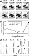
Figure 1
Figure 1. Naive antiviral CD4+ and CD8+ T cell division has a lag phase of 2–3 days.
Equal numbers of CFSE-labeled P14 cells (TcR-transgenic CD8+ T cells specific for LCMV GP33–41, expressing Thy1.1) and CFSE-labeled SMARTA cells (TcR transgenic CD4+ T cells specific for LCMV GP61–80, expressing Ly5a) were pooled, and inoculated into wildtype C57BL/6 mice, which then were infected with LCMV. A. At the indicated times after infection, each donor population was identified by flow cytometry (ovals). B. The numbers of P14 and SMARTA T cells in the spleen are shown (mean±SE) at the indicated times after infection (two separate experiments, two mice per experiment). C. After gating to identify the P14 or the SMARTA T cells, the histograms show these cells' CFSE fluorescence. Note that both T cell subsets begin proliferating at the same time (day 3).
Figure 2
Figure 2. The delay in naïve T cell division is organism-wide.
Mice containing approximately 1.4×10⁵ CFSE-labeled SMARTA CD4+ T cells were infected with LCMV. At the indicated times after infection, lymphocytes were isolated (2 mice per time point) and the donor cells were identified by flow cytometry. A. The ovals in the dot plots identify the SMARTA CD4+ T cells, and the numbers indicate their percentage among leukocytes isolated from each tissue. The histograms show the CFSE-fluorescence of the SMARTA CD4+ T cells; the numbers indicate the percentages of SMARTA CD4+ T cells that have divided. B. The line graphs show the percentages of SMARTA CD4+ T cells among all isolated leukocytes at various times after infection. For each tissue, the dashed line indicates the number of SMARTA cells in uninfected mice.
Figure 3
Figure 3. Kinetics of naive and memory CD4+ T cells in the same mouse.
Wildtype mice containing 1.3×10³ naïve SMARTA (Thy1.1) and 1.3×10³ memory SMARTA (Ly5a) cells were given LCMV, and the relative abundance of the two SMARTA cell populations was determined by flow cytometry at various times post infection (two mice per time point). A. After gating on CD4+ T cells, the host CD4+ T cells (H), and the naïve and memory SMARTA cells (N & M respectively) were distinguished by Thy1.1 and Ly5a staining. The numbers indicate the frequencies of naïve and memory SMARTA cells as a percentage of all CD4+ T cells. B. The average±SE of the percentage of each population among all CD4+ T cells is shown over time. C. The total number of memory or naïve SMARTA CD4+ T cells per spleen is shown (average±SE).
Figure 4
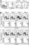
Figure 4. Naive and memory CD4+ T cells show near-identical delays in onset of division.
Mice containing 2×10³ naive SMARTA CD4+ T cells (Ly5a) were infected with LCMV and allowed to become immune. A. Six months after infection, memory SMARTA CD4+ T cells were isolated from the spleen and analyzed by flow cytometry. The first dot plot identifies the memory SMARTA CD4+ T cells (oval). After gating on these cells, the histogram shows their expression of CD44, and the remaining two dot plots evaluate IFNγ and IL-2 production after brief in vitro stimulation with GP61–80 peptide. B. The memory SMARTA cells (Ly5a) were mixed with naive SMARTA cells (Thy1.1), labeled with CFSE, and then transferred to naive mice. The recipient mice were given approximately 5×10⁴ memory SMARTA CD4 T cells and 5×10⁵ naive SMARTA CD4 T cells. 3 days later, the recipients were infected with LCMV. The dot plots show spleen cells isolated from recipient mice at the indicated times after infection, and the ovals identify the memory SMARTA CD4 T cells (top two rows) and the naive SMARTA CD4 T cells (bottom two rows). The histograms show the CFSE fluorescence of the SMARTA cells, and the numbers in the histograms indicate the percentage of SMARTA CD4+ T cells that have not divided. Data are representative of two independent experiments.
Figure 5
Figure 5. Delayed accumulation of naive and memory CD4+ T cells occurs also in non-lymphoid tissues.
Mice containing 1.3×10⁴ naïve (Thy1.1) and 1.3×10⁴ memory (Ly5a) SMARTA CD4+ T cells were infected with LCMV and, at the indicated times after infection, lymphocytes from several lymphoid and non-lymphoid tissues were isolated and analyzed by flow cytometry. A. Dot plots show gated CD4+ T cells isolated from the tissues. The ovals identify the SMARTA cells (N, M  =  naïve & memory respectively), and the numbers indicate their percentage among all CD4 T cells (H  =  host CD4+ T cells). B. For each tissue, naïve and memory SMARTA CD4+ T cells are shown as percentages of all CD4+ T cells (two mice per time point). Note that both naïve and memory cells become prominent after day 4; however, the memory cells dominate the response in the non-lymphoid tissues.
Figure 6
Figure 6. Viral epitopes are presented within hours of infection, and stimulate memory T cell effector functions.
Mice that contained approximately 3×10³ SMARTA/Ly5a CD4+ T cells were infected with LCMV and, 354 days later, were re-challenged intraperitoneally with 2×10⁶ PFU LCMV-Armstong. Six hours post-infection, the mice were given 0.25 mg Brefeldin A i.v., and 6 hours later the spleens were harvested and immediately surface stained for CD4, Ly5a, or CD8, then permeabilized and stained for intracellular IFNγ. The cells were not re-stimulated ex vivo with peptide antigen. A. ∼5% of all CD8+ T cells, and ∼1% of all CD4+ T cells, are actively producing IFNγ in response to infection. B. Using the SMARTA cells transferred ∼1 year previously as an indicator of the responsiveness of virus-specific CD4+ memory T cells, ∼14% of LCMV-specific CD4+ memory T cells actively produce IFNγ within 12 hours of virus infection. Data shown are from an individual mouse, and are representative of independent datasets. C. A separate set of naive mice were given CFSE-labeled pooled SMARTA cells (4×10⁵ naive SMARTA/Thy1.1 cells and 2×10⁴ memory SMARTA/Ly5a T cells). 4 days later, some of the recipient mice were given LCMV. Six hours later, BFA was administered to all mice, and after a further 6 hours splenocytes were harvested. The cells were immediately stained (without peptide re-stimulation) for CD4, Thy1.1, Ly5a and IFNγ, and were analyzed by flow cytometry. Approximately 2% memory SMARTA cells had begun to synthesize IFNγ in response to LCMV infection (top row) but none of those responding memory cells showed any dilution of CFSE signal. The naïve SMARTA cells (bottom row) failed to produce IFNγ at this early time point post-infection, and no sign of cell division was seen. Data are from one of two independent experiments.
Figure 7
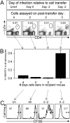
Figure 7. Changing the microenvironment reduces the in vivo delay in T cell division.
Naive SMARTA cells were CFSE-labeled and transferred either to mice that had been infected with LCMV two days previously, or to uninfected mice some of which were immediately infected with LCMV. A. 2, 3 or 4 days after cell transfer (as indicated), the spleens of the recipient mice were isolated and the donor SMARTA CD4+ T cells were identified by flow cytometry (ovals). Individual mice are shown, and the numbers indicate the proportion of SMARTA cells as a percentage of all spleen cells. Mouse numbers in each of the 4 groups: 1, 1, 3, 3. B. The bar graph shows cumulative data, as percentages of SMARTA CD4+ T cells. C. The histograms show the CFSE fluorescence of the indicated SMARTA CD4+ T cells. Note that the 3-day delay in proliferation is shortened to 2 days if the mice were pre-infected.

Similar articles

Cited by

References

    1. Veiga-Fernandes H, Walter U, Bourgeois C, McLean A, Rocha B. Response of naive and memory CD8+ T cells to antigen stimulation in vivo. Nat Immunol. 2000;1:47–53. - PubMed
    1. Rogers PR, Dubey C, Swain SL. Qualitative changes accompany memory T cell generation: faster, more effective responses at lower doses of antigen. J Immunol. 2000;164:2338–2346. - PubMed
    1. Slifka MK, Whitton JL. Functional avidity maturation of CD8+ T cells without selection of higher affinity TCR. Nat Immunol. 2001;2:711–717. - PubMed
    1. Lefrancois L. Development, trafficking, and function of memory T-cell subsets. Immunol Rev. 2006;211:93–103. - PubMed
    1. Weninger W, Crowley MA, Manjunath N, von Andrian UH. Migratory properties of naive, effector, and memory CD8+ T cells. J Exp Med. 2001;194:953–966. - PMC - PubMed

Publication types

MeSH terms