Review
. 2008 Sep;37(7):1271-8.
doi: 10.1007/s00249-008-0321-0. Epub 2008 Apr 11.

Push-me-pull-you: how microtubules organize the cell interior

Affiliations
Review

Push-me-pull-you: how microtubules organize the cell interior

Iva M Tolić-Nørrelykke. Eur Biophys J. 2008 Sep.

Abstract

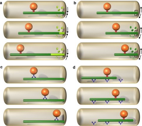
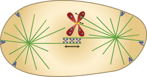
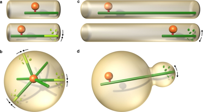
Dynamic organization of the cell interior, which is crucial for cell function, largely depends on the microtubule cytoskeleton. Microtubules move and position organelles by pushing, pulling, or sliding. Pushing forces can be generated by microtubule polymerization, whereas pulling typically involves microtubule depolymerization or molecular motors, or both. Sliding between a microtubule and another microtubule, an organelle, or the cell cortex is also powered by molecular motors. Although numerous examples of microtubule-based pushing and pulling in living cells have been observed, it is not clear why different cell types and processes employ different mechanisms. This review introduces a classification of microtubule-based positioning strategies and discusses the efficacy of pushing and pulling. The positioning mechanisms based on microtubule pushing are efficient for movements over small distances, and for centering of organelles in symmetric geometries. Mechanisms based on pulling, on the other hand, are typically more elaborate, but are necessary when the distances to be covered by the organelles are large, and when the geometry is asymmetric and complex. Thus, taking into account cell geometry and the length scale of the movements helps to identify general principles of the intracellular layout based on microtubule forces.


Figures

Fig. 1
Fig. 1
Basic types of microtubule force generation. a Pushing, b pulling; c, d sliding. a, b The organelle (orange) is bound to the microtubule (green) by a fixed link (red). a The organelle is being pushed away from the cell edge by a microtubule polymerization force. The microtubule polymerizes by addition of new subunits (light green discs) at its end (arrows). b A depolymerizing microtubule, which is connected to the cell edge by an active motor or a passive “adaptor” (dark grey), is pulling the organelle towards the cell edge. Depolymerization is accompanied by a loss of old subunits (dark green discs and arrows). c A motor protein (blue) walks along the microtubule and carries the organelle. d Motor proteins (blue) are anchored at the cell cortex and walk along the microtubule, thus translating the microtubule together with the bound organelle
Fig. 2
Fig. 2
The mitotic spindle. Microtubules (green) form two asters and the central spindle, where the microtubules growing from the two spindle poles meet. Microtubule “plus-ends” (the more dynamic ends) are in the center of the spindle and at the cell periphery, while the “minus-ends” (the less dynamic ends) are anchored at the spindle poles. Motor proteins (blue, at the cell center) cross-link the overlapping anti-parallel microtubules in the central spindle and slide them past each other. Chromosomes (red; only one is shown for clarity) are attached to microtubules at the kinetochore (yellow), where forces towards and away from the spindle pole are generated. Chromokinesins (grey) reside at the chromosome arms and push the chromosomes away from the spindle pole by walking towards the microtubule plus-end. Minus-end directed motors (blue, at the cell periphery) are anchored at the cell cortex and pull on astral microtubules, thereby elongating the spindle
Fig. 3
Fig. 3
Pushing versus pulling. The positioning mechanisms based on microtubule pushing are efficient for movements over small distances (a), and for centering in symmetric geometries (b). Mechanisms based on pulling are preferred in cases where the distances to be covered by the organelles are large (c), and where the geometry is asymmetric and complex (d)

References

    1. Adames NR, Cooper JA. Microtubule interactions with the cell cortex causing nuclear movements in Saccharomyces cerevisiae. J Cell Biol. 2000;149:863–874. doi: 10.1083/jcb.149.4.863. - DOI - PMC - PubMed
    1. Aist JR, Berns MW. Mechanics of chromosome separation during mitosis in Fusarium (Fungi imperfecti): new evidence from ultrastructural and laser microbeam experiments. J Cell Biol. 1981;91:446–458. doi: 10.1083/jcb.91.2.446. - DOI - PMC - PubMed
    1. Aist JR, Liang H, Berns MW. Astral and spindle forces in PtK2 cells during anaphase B: a laser microbeam study. J Cell Sci. 1993;104:1207–1216. - PubMed
    1. Alberts B, Johnson A, Lewis J, Raff M, Roberts K, Walter P. Molecular biology of the cell. 5th edn. New York: Garland Science; 2008.
    1. Antonio C, Ferby I, Wilhelm H, Jones M, Karsenti E, Nebreda AR, Vernos I. Xkid, a chromokinesin required for chromosome alignment on the metaphase plate. Cell. 2000;102:425–435. doi: 10.1016/S0092-8674(00)00048-9. - DOI - PubMed

LinkOut - more resources