. 2007 Mar;3(1-2):13-25.
doi: 10.1007/s11302-006-9033-z. Epub 2007 Jan 30.

P1 receptors and cytokine secretion

Affiliations

P1 receptors and cytokine secretion

Maria P Abbracchio et al. Purinergic Signal. 2007 Mar.

Abstract

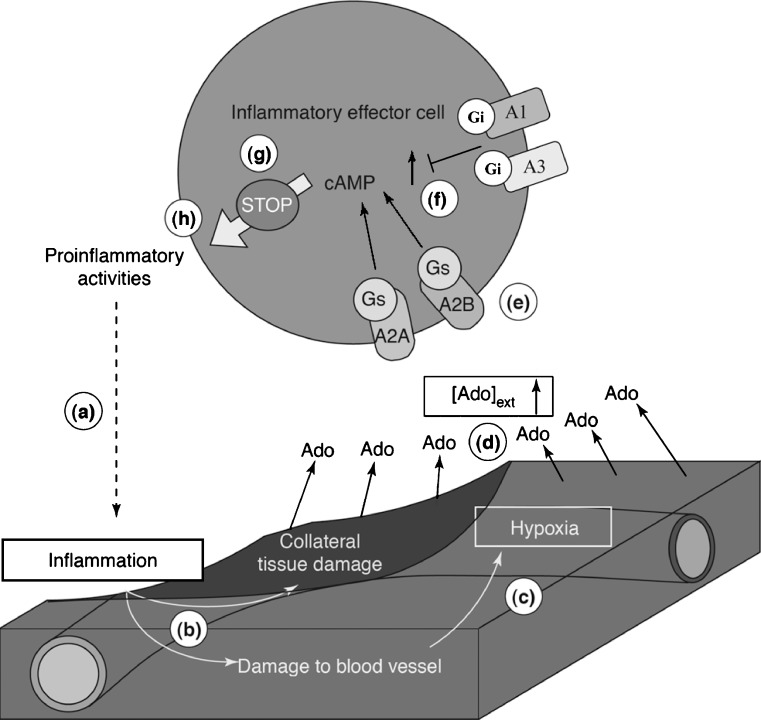
Evidence has accumulated in the last three decades to suggest tissue protection and regeneration by adenosine in multiple different cell types. Adenosine produced in hypoxic or inflamed environments reduces tissue injury and promotes repair by receptor-mediated mechanisms. Among other actions, regulation of cytokine production and secretion by immune cells, astrocytes and microglia (the brain immunocytes) has emerged as a main mechanism at the basis of adenosine effects in diseases characterized by a marked inflammatory component. Many recent studies have highlighted that signalling through A(1) and A(2A) adenosine receptors can powerfully prevent the release of pro-inflammatory cytokines, thus inhibiting inflammation and reperfusion injury. However, the activation of adenosine receptors is not invariably protective of tissues, as signalling through the A(2B) adenosine receptor has been linked to pro-inflammatory actions which are, at least in part, mediated by increased release of pro-inflammatory cytokines from epithelial cells, astrocytes and fibroblasts. Here, we discuss the multiple actions of P1 receptors on cytokine secretion, by analyzing, in particular, the role of the various adenosine receptor subtypes, the complex reciprocal interplay between the adenosine and the cytokine systems, their pathophysiological significance and the potential of adenosine receptor ligands as new anti-inflammatory agents.


Figures

Fig. 1
Fig. 1
Role of A2 adenosine receptors in delayed negative feedback downregulation of activated immune cells in inflammation. a Pathogens, virus-infected, mutated or otherwise injured cells activate resident, recruited or tissue-surveying immune cells inducing them to produce pro-inflammatory molecules (e.g. cytokines, reactive oxygen species). b This leads to pathogen destruction and, as a result of strong activation, some non-infected, ‘innocent–bystander cells in collateral tissue can also be damaged. c According to the hypothesis raised by Sitkovsky and Ohta [10], it is the collateral damage to the microvasculature owing to the continuing production of inflammatory molecules that is of crucial significance in signalling the extent of tissue damage. In particular, increased damage to microvasculature may result in the interruption of blood supply and low oxygen tension (hypoxia) in the most damaged microenvironment. d Local tissue hypoxia leads to the accumulation of extracellular adenosine (Ado), as a result of both hypoxia-associated decrease in intracellular levels of ATP and inhibition of adenosine kinase (see text). e As the oxygen tension falls further, the concentrations of extracellular adenosine ([Ado]ext) will increase and this, in turn, will determine the intensity of signalling through sequential recruitment of high-affinity (A2A) and low-affinity (A2B) adenosine receptors. f The sufficiently high extracellular adenosine levels will trigger the maximal activation of Gs protein-coupled A2A and A2B adenosine receptors and the accumulation of intracellular cAMP, which has strong immunosuppressive properties. g The increased cAMP then strongly inhibits ongoing effector functions and prevents their triggering in the newly activated immune cells that have just arrived in the inflamed area. Immune cells also express Gi protein-coupled A1 and A3 receptors, which inhibit adenylyl cyclase and cAMP formation, which, in turn, would provide another level of control to prevent the premature inhibition of immune cells by A2 receptors. h This delayed negative feedback mechanism might provide immune cells sufficient time to destroy the pathogen but also prevents additional collateral tissue damage by inhibiting the production of pro-inflammatory cytokines and cytotoxic molecules. Reprinted and modified from Sitkovsky and Ohta [10], copyright 2005 with permission from Elsevier
Fig. 2
Fig. 2
Role of A2 adenosine receptors in the modulation of the release of histamine in the lung under physiological or pathological conditions. Both high-affinity A2A and low-affinity A2B adenosine receptors are positively coupled to cAMP production through Gs. In addition, the A2B subtype can also promote Ins(1,4,5)P3 production via Gq activation. Under normal conditions a, low extracellular adenosine concentrations activate the A2A receptor subtype, leading to the increase of intracellular cAMP concentrations, which are known to inhibit histamine release. In asthma and COPD, high extracellular adenosine concentrations are reached b. This in turn might lead to the downregulation of high-affinity A2A receptors and might therefore increase the relative importance of the low-affinity A2B subtype. The concomitant reduction in cAMP concentrations, paralleled by overproduction of Ins(1,4,5)P3 through Gq activation, will promote histamine release as the final outcome, thus contributing to the development and exacerbation of the disease. Reprinted from Spicuzza et al. [27], copyright 2003 with permission from Elsevier
Fig. 3
Fig. 3
In vitro induction of reactive astrogliosis by the A2B adenosine receptor in TNFα-treated cells. Exposure of human astrocytoma cells to TNFα increases A2B receptor signalling and G protein coupling by reducing agonist-dependent receptor phosphorylation and desensitization, without affecting receptor protein and mRNA levels (not shown; see Trincavelli et al. [68] for details). From a functional point of view, these biochemical changes translate into the ability of the A2B receptor to induce elongation of astrocytic processes, a typical hallmark of reactive astrogliosis. In fact, marked morphological changes can be observed in cells preincubated with 1,000 U/ml TNFα for 24 h, and subsequently exposed to 1 μM NECA for 30 min, followed by an additional 72 h in drug-free medium b, with respect to cultures exposed to TNFα alone, which induced no effect ‘per se–a. Quantification of results indicates a 25-30% elongation of cell processes by NECA in the presence of TNFα, with respect to TNFα alone. No effect on process elongation was detected when cells were exposed to NECA without TNFα pretreatment (see Trincavelli et al. [68] for details). NECA-induced astrocytic elongation can be completely inhibited by the concomitant exposure to MRS 1706 10 nM, c, a selective A2B antagonist, thus confirming a specific involvement of this receptor subtype in the observed effects. Magnification: ×32. Scale bar: 30 μM. Reprinted from Trincavelli et al. [68], copyright 2004 with permission from Blackwell Publishing
Fig. 4
Fig. 4
Alterations of peripheral A2A receptors in CHF patients progressively normalize after heart transplantation in parallel with the normalization of haemodynamic conditions and of plasma cytokine concentrations. In CHF patients, plasma levels of adenosine, TNFα and its soluble receptor are elevated, whereas in peripheral circulating cells A2A adenosine receptors are upregulated (see Varani et al. [80] for more details). In these patients, the latter change mirrors the A2A receptor changes occurring in the heart, the disease target organ. In a cohort of these patients, A2A receptors in peripheral blood circulating cells have been studied longitudinally as a function of time before and after heart transplantation. The graph shows the longitudinal analysis of A2A receptor density (Bmax) as determined with [3H]-ZM 241385 binding in the lymphocytes of six patients, before and at different times after heart transplantation (see inset for legend to individual cases). Dotted horizontal line indicates the mean value of A2A receptor density in control healthy subjects. Receptor density gradually returned to normal values within 6 months after transplant. Similarly, the KD value of [3H]-ZM 241385 binding gradually and progressively decreased to control values within the same time period (data not shown). This trend was consistently evident for all evaluated patients and was also detected in the neutrophils of the same subjects, showing a progressive normalization of binding parameters to control values as a function of time. In transplanted patients, plasma adenosine TNFα and IL-6 levels also showed a trend to a decrease to values within 3-6 months after transplant (see Capecchi et al. [82]). Therefore, the cytokine milieu may regulate the function and the expression of A2A adenosine receptors, thus contributing to establishing a negative feedback mechanism against the progressive loop between pro-inflammatory cytokines and heart failure (see also Capecchi et al. [82]). Transplantation results in normalization of haemodynamics, reduction of inflammation and normalization of the number and function of A2A adenosine receptors. Modified from Varani et al. [80], copyright 2003 with permission from The Federation of American Society for Experimental Biology

References

    1. Jacobson KA, Gao ZG. Adenosine receptors as therapeutic targets. Nat Rev Drug Discov. 2006;5:247–264. doi: 10.1038/nrd1983. - DOI - PMC - PubMed
    1. Linden J. Adenosine in tissue protection and tissue regeneration. Mol Pharmacol. 2005;67:1406–1413. doi: 10.1124/mol.105.011783. - DOI - PubMed
    1. Hirschhorn R, Grossman J, Weissmann G. Effect of cyclic 3′5′-adenosine monophosphate and theophylline on lymphocyte transformation. Proc Soc Exp Biol Med. 1970;133:1361–1365. - PubMed
    1. Henney CS, Lichtenstein LM. The role of cyclic AMP in the cytolytic activity of lymphocytes. J Immunol. 1971;107:610–612. - PubMed
    1. Cronstein BN, Kramer SB, Rosenstein ED, et al. Adenosine modulates the generation of superoxide anion by stimulated human neutrophils via interaction with a specific cell surface receptor. Ann NY Acad Sci. 1985;451:291–301. doi: 10.1111/j.1749-6632.1985.tb27120.x. - DOI - PubMed

LinkOut - more resources