. 2007 Mar;3(1-2):71-81.
doi: 10.1007/s11302-006-9038-7. Epub 2007 Jan 9.

Extracellular NAD and ATP: Partners in immune cell modulation

Affiliations

Extracellular NAD and ATP: Partners in immune cell modulation

Friedrich Haag et al. Purinergic Signal. 2007 Mar.

Abstract

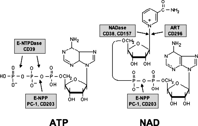
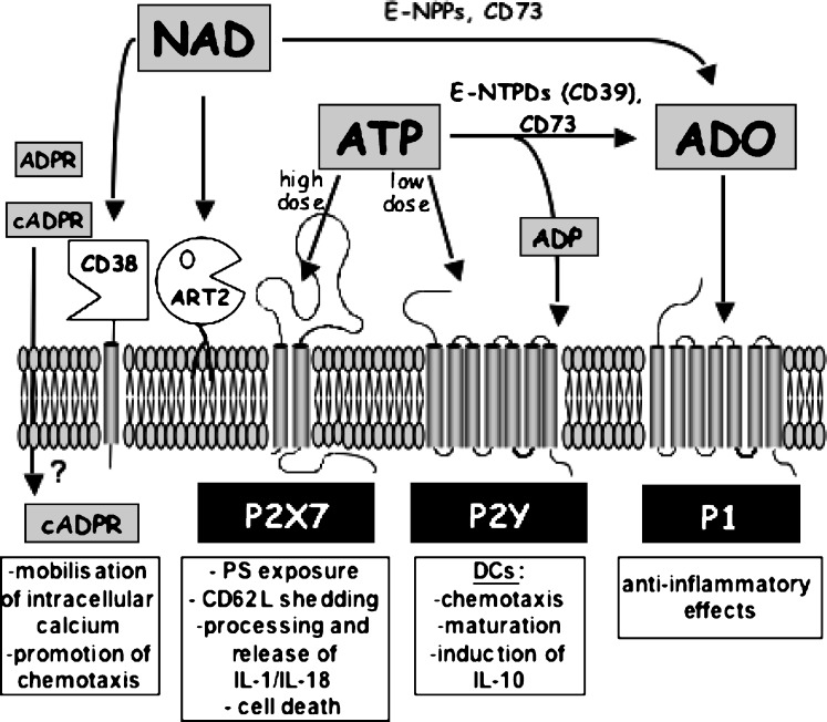
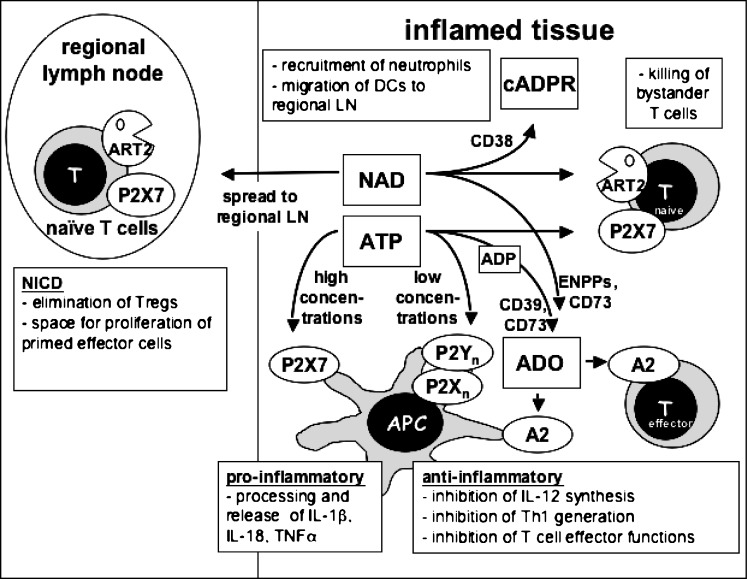
Extracellular NAD and ATP exert multiple, partially overlapping effects on immune cells. Catabolism of both nucleotides by extracellular enzymes keeps extracellular concentrations low under steady-state conditions and generates metabolites that are themselves signal transducers. ATP and its metabolites signal through purinergic P2 and P1 receptors, whereas extracellular NAD exerts its effects by serving as a substrate for ADP-ribosyltransferases (ARTs) and NAD glycohydrolases/ADPR cyclases like CD38 and CD157. Both nucleotides activate the P2X7 purinoceptor, although by different mechanisms and with different characteristics. While ATP activates P2X7 directly as a soluble ligand, activation via NAD occurs by ART-dependent ADP-ribosylation of cell surface proteins, providing an immobilised ligand. P2X7 activation by either route leads to phosphatidylserine exposure, shedding of CD62L, and ultimately to cell death. Activation by ATP requires high micromolar concentrations of nucleotide and is readily reversible, whereas NAD-dependent stimulation begins at low micromolar concentrations and is more stable. Under conditions of cell stress or inflammation, ATP and NAD are released into the extracellular space from intracellular stores by lytic and non-lytic mechanisms, and may serve as "danger signals" to alert the immune response to tissue damage. Since ART expression is limited to naïve/resting T cells, P2X7-mediated NAD-induced cell death (NICD) specifically targets this cell population. In inflamed tissue, NICD may inhibit bystander activation of unprimed T cells, reducing the risk of autoimmunity. In draining lymph nodes, NICD may eliminate regulatory T cells or provide space for the preferential expansion of primed cells, and thus help to augment an immune response.


Figures

Figure 1
Figure 1
Chemical structure of ATP and NAD, and sites of cleavage by different ecto-enzymes
Figure 2
Figure 2
Action of extracellular ATP and NAD and their metabolites on different cell surface receptors. Extracellular ATP present in high, intermediate, or low concentrations can activate P2X7, other P2X, or P2Y receptors, respectively, or is hydrolysed by the sequential action of ecto-nucleoside triphosphate diphosphohydrolases (E-NTPDs) such as CD39 and ecto-5–nucleotidase (CD73) to ADP and adenosine (ADO). For clarity, P2X receptors other than P2X7 are not shown, since their presence on immune cells is not well documented. ADP can act on P2Y receptors, and adenosine can activate G protein-coupled P1 receptors. Extracellular NAD serves as a substrate for cell-surface ADP-ribosyltransferases (ART2), or is hydrolyzed to ADPribose by CD38. CD38 can also synthesise cyclic ADP-ribose, a known intracellular calcium mobilising agent. It is not known how cADPR gains access to the intracellular compartment. NAD (and ATP) may also be hydrolysed by ecto-nucleotide pyrophosphatase/phosphodiesterases (E-NPPs) to AMP, which in turn is hydrolysed by CD73 to adenosine. See text for details
Figure 3
Figure 3
Hypothetical scheme of the interplay of purine sensors during an immune response. ATP and NAD are released locally at sites of tissue injury or inflammation. At high concentrations, ATP acts on the P2X7R receptor to exert pro-inflammatory effects on antigen presenting cells or to kill T cells; at low concentrations it acts on other P2 receptors to downregulate the initiation of Th1 responses. NAD is used by ARTs on T cells to activate P2X7, or by CD38 to generate cyclic ADP-ribose. It is conceivable that NAD may exert distant effects by reaching lymph nodes draining inflammatory sites in physiologically relevant concentrations. Both ATP and NAD are degraded by metabolising enyzmes to yield other signalling molecules, notably adenosine (ADO), which exerts predominantly anti-inflammatory effects through P1 receptors of the A2-subfamily. See text for details

References

    1. Berger F, Lau C, Dahlmann M, et al. Subcellular compartmentation and differential catalytic properties of the three human nicotinamide mononucleotide adenylyltransferase isoforms. J Biol Chem. 2005;280:36334–36341. doi: 10.1074/jbc.M508660200. - DOI - PubMed
    1. D’Amours D, Desnoyers S, D’Silva I, et al. Poly(ADP-ribosyl)ation reactions in the regulation of nuclear functions. Biochem J. 1999;342:249–268. doi: 10.1042/0264-6021:3420249. - DOI - PMC - PubMed
    1. Herceg Z, Wang ZQ. Functions of poly(ADP-ribose) polymerase (PARP) in DNA repair, genomic integrity and cell death. Mutat Res. 2001;477:97–110. - PubMed
    1. Di Lisa F, Ziegler M. Pathophysiological relevance of mitochondria in NAD(+) metabolism. FEBS Lett. 2001;492:4–8. doi: 10.1016/S0014-5793(01)02198-6. - DOI - PubMed
    1. Schwiebert EM, Zsembery A. Extracellular ATP as a signaling molecule for epithelial cells. Biochim Biophys Acta. 2003;1615:7–32. doi: 10.1016/S0005-2736(03)00210-4. - DOI - PubMed