. 2007 Mar;3(1-2):171-80.
doi: 10.1007/s11302-006-9050-y. Epub 2007 Feb 6.

CD39 and control of cellular immune responses

Affiliations

CD39 and control of cellular immune responses

Karen M Dwyer et al. Purinergic Signal. 2007 Mar.

Abstract

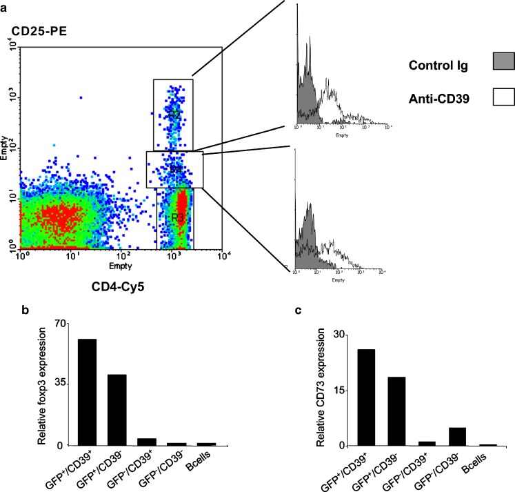
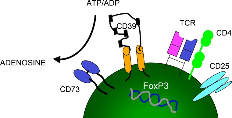
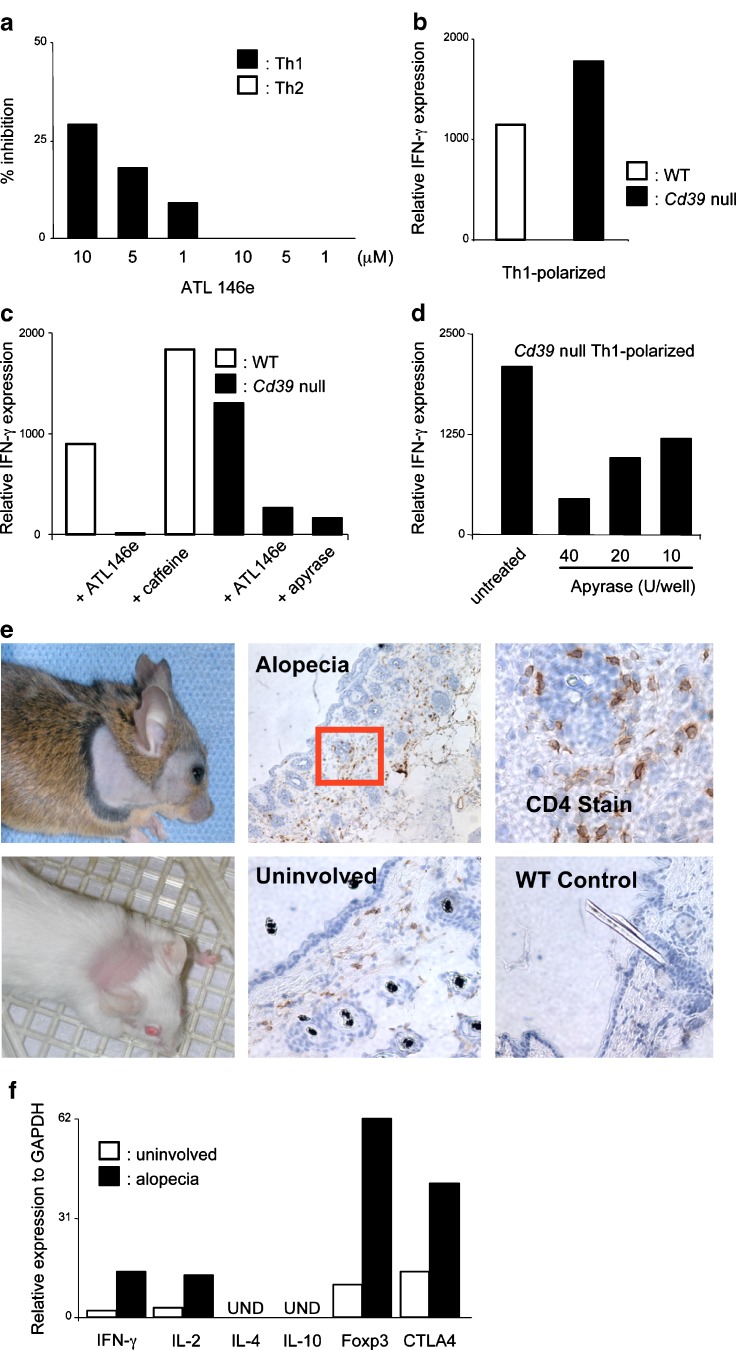
CD39 is the cell surface-located prototypic member of the ecto-nucleoside triphosphate diphosphohydrolase (E-NTPDase) family. Biological actions of CD39 are a consequence (at least in part) of the regulated phosphohydrolytic activity on extracellular nucleotides. This ecto-enzymatic cascade in tandem with CD73 (ecto-5'-nucleotidase) also generates adenosine and has major effects on both P2 and adenosine receptor signalling. Despite the early recognition of CD39 as a B lymphocyte activation marker, little is known of the role of CD39 in humoral or cellular immune responses. There is preliminary evidence to suggest that CD39 may impact upon antibody affinity maturation. Pericellular nucleotide/nucleoside fluxes caused by dendritic cell expressed CD39 are also involved in the recruitment, activation and polarization of naïve T cells. We have recently explored the patterns of CD39 expression and the functional role of this ecto-nucleotidase within quiescent and activated T cell subsets. Our data indicate that CD39, together with CD73, efficiently distinguishes T regulatory cells (Treg) from other resting or activated T cells in mice (and humans). Furthermore, CD39 serves as an integral component of the suppressive machinery of Treg, acting, at least in part, through the modulation of pericellular levels of adenosine. We have also shown that the coordinated regulation of CD39/CD73 expression and of the adenosine receptor A2A activates an immunoinhibitory loop that differentially regulates Th1 and Th2 responses. The in vivo relevance of this network is manifest in the phenotype of Cd39-null mice that spontaneously develop features of autoimmune diseases associated with Th1 immune deviation. These data indicate the potential of CD39 and modulated purinergic signalling in the co-ordination of immunoregulatory functions of dendritic and Treg cells. Our findings also suggest novel therapeutic strategies for immune-mediated diseases.


Figures

Fig. 1
Fig. 1
a Expression of CD39 on mouse lymphoid cells. Lymph node cell suspensions were prepared from 8-week-old C57/BL6 mice and were gated based on FSC and SSC parameters (not shown). CD4+ cells were gated based on CD25 expression. CD39 expression on the different subsets is shown in the histograms (open profiles) plotted against an irrelevant control (gray profiles). Differential expression of CD39 on CD4+ cells can be shown to be closely associated with CD25. b RT-PCR analysis of foxp3 mRNA expression. This was done in sorted cells with the following markers: Foxp3+/CD39+, Foxp3+/CD39-, Foxp3-/CD39+ and Foxp3-/CD39-. CD4+ cells obtained from the Foxp3-GFP ‘knockin–animals that had been generated by Mohamed Oukka and Vijay K. Kuchroo with colleagues (Ref). B lymphocytes were used as CD39+ control and included for comparison (striped bars). c RT-PCR analysis of CD73 mRNA expression. This was done in sorted cells with the following markers: Foxp3+/CD39+, Foxp3+/CD39-, Foxp3-/CD39+ and Foxp3-/CD39-. B lymphocytes were used as CD39+ control and included for comparison (striped bars). CD73 is also a useful immunophenotypic marker for Treg cells (not shown); and when combined with CD39 provides near-concordance with foxp3 expression. The Foxp3-/CD39+ subset does not express CD73 and these cells resemble the memory phenotype (not shown)
Fig. 2
Fig. 2
Schematic representation of Treg markers. The cellular phenotype of these suppressive T cells can be defined by FoxP3+/CD39+/CD73+ expression. Phosphohydrolysis of extracellular nucleotides by CD39 and CD73 generates adenosine, which exerts a component of the immunosuppressive effect
Fig. 3
Fig. 3
CD39 generation of adenosine preferentially regulates Th1 immune responses. a 3H-thymidine incorporation of CD4+ T cells purified from WT mice as polarized towards a Th1 or a Th2 phenotype for 5 days. The selective adenosine A2A agonist ATL146e was added after 3 days of culture. Data are expressed as % of inhibition and are the mean of duplicates; error bars represent the SEM of three independent experiments. b CD4+ T cells were purified from Cd39-null (filled histograms) or WT (open histograms) mice and polarized towards a Th1 phenotype. The mRNA was extracted at day 3 and assayed for IFN-g (left panel) by RT-PCR. Data are representative of more than four independent experiments. c CD4+ T cells were purified from Cd39-null (filled histograms) or WT (open histograms) mice and polarized towards a Th1 phenotype. The indicated adenosine receptor agonists and antagonists were added at the beginning of the experiment (left panel). d This panel shows the effects of apyrase on IFN-g production by Cd39-null CD4+ T cells polarized towards a Th1 phenotype. For these experiments, mRNA was extracted at day 3 and assayed for IFN-g (RT-PCR). Representative data are shown from three independent experiments for each. e Representative images of Cd39-null mouse on C57BL/6/129 SVJ (upper panel) or BALB/c backgrounds (lower panel) manifesting alopecia. Histology of skin samples obtained from Cd39-null mice affected by alopecia. Uninvolved skin areas from the same animals as well as samples from WT mice were used for comparison. Biopsies were fixed and stained for CD4 cells. Original magnifications: ×10 for the upper middle panel, ×40 for the upper right and ×20 for the lower panels. f Skin samples were obtained from Cd39-null mice affected by alopecia, the tissue homogenized and mRNA extracted. Cytokine profiling was performed by RT-PCR, using a preamplification technique. Uninvolved skin from the same animal or a matched WT mouse used as controls. Representative data are from three animals

References

    1. Robson SC, Wu Y, Sun X, Knosalla C, Dwyer K, Enjyoji K. Ectonucleotidases of CD39 family modulate vascular inflammation and thrombosis in transplantation. Semin Thromb Hemost. 2005;31:217–233. doi: 10.1055/s-2005-869527. - DOI - PubMed
    1. Robson SC, Sevigny J, Zimmermann H. The E-NTPDase family of ectonucleotidases: structure function relationships and pathophysiological significance. Purinergic Signalling. 2006;2:409–430. doi: 10.1007/s11302-006-9003-5. - DOI - PMC - PubMed
    1. Luthje J. Origin, metabolism and function of extracellular adenine nucleotides in the blood [published erratum appears in Klin Wochenschr (1989) 67(10):558] Klin Wochenschr. 1989;67:317–327. doi: 10.1007/BF01741386. - DOI - PubMed
    1. la Sala A, Ferrari D, Di Virgilio F, Idzko M, Norgauer J, Girolomoni G. Alerting and tuning the immune response by extracellular nucleotides. J Leukoc Biol. 2003;73:339–343. doi: 10.1189/jlb.0802418. - DOI - PubMed
    1. Imai M, Goepfert C, Kaczmarek E, Robson SC. CD39 modulates IL-1 release from activated endothelial cells. Biochem Biophys Res Commun. 2000;270:272–278. doi: 10.1006/bbrc.2000.2410. - DOI - PubMed