Mapping P2X and P2Y receptor proteins in striatum and substantia nigra: An immunohistological study
- PMID: 18404452
- PMCID: PMC2072921
- DOI: 10.1007/s11302-007-9069-8
Mapping P2X and P2Y receptor proteins in striatum and substantia nigra: An immunohistological study
Abstract
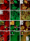
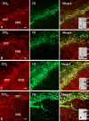
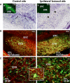
Our work aimed to provide a topographical analysis of all known ionotropic P2X(1-7) and metabotropic P2Y(1,2,4,6,11-14) receptors that are present in vivo at the protein level in the basal ganglia nuclei and particularly in rat brain slices from striatum and substantia nigra. By immunohistochemistry-confocal and Western blotting techniques, we show that, with the exception of P2Y(11,13) receptors, all other subtypes are specifically expressed in these areas in different amounts, with ratings of low (P2X(5,6) and P2Y(1,6,14) in striatum), medium (P2X(3) in striatum and substantia nigra, P2X(6,7) and P2Y(1) in substantia nigra) and high. Moreover, we describe that P2 receptors are localized on neurons (colocalizing with neurofilament light, medium and heavy chains) with features that are either dopaminergic (colocalizing with tyrosine hydroxylase) or GABAergic (colocalizing with parvalbumin and calbindin), and they are also present on astrocytes (P2Y(2,4), colocalizing with glial fibrillary acidic protein). In addition, we aimed to investigate the expression of P2 receptors after dopamine denervation, obtained by using unilateral injection of 6-hydroxydopamine as an animal model of Parkinson's disease. This generates a rearrangement of P2 proteins: most P2X and P2Y receptors are decreased on GABAergic and dopaminergic neurons, in the lesioned striatum and substantia nigra, respectively, as a consequence of dopaminergic denervation and/or neuronal degeneration. Conversely, P2X(1,3,4,6) on GABAergic neurons and P2Y(4) on astrocytes augment their expression exclusively in the lesioned substantia nigra reticulata, probably as a compensatory reaction to dopamine shortage. These results disclose the presence of P2 receptors in the normal and lesioned nigro-striatal circuit, and suggest their potential participation in the mechanisms of Parkinson's disease.
Figures


Similar articles
-
P2 receptor expression in the dopaminergic system of the rat brain during development.Neuroscience. 2007 Oct 12;149(1):165-81. doi: 10.1016/j.neuroscience.2007.07.015. Epub 2007 Jul 17. Neuroscience. 2007. PMID: 17869006
-
Altered melatonin MT1 receptor expression in the ventral midbrain following 6-hydroxydopamine lesions in the rat medial forebrain bundle.Brain Res. 2016 Dec 1;1652:89-96. doi: 10.1016/j.brainres.2016.09.036. Epub 2016 Sep 28. Brain Res. 2016. PMID: 27693415
-
Downregulation of glial glutamate transporters after dopamine denervation in the striatum of 6-hydroxydopamine-lesioned rats.J Comp Neurol. 2008 Dec 1;511(4):421-37. doi: 10.1002/cne.21852. J Comp Neurol. 2008. PMID: 18831527
-
P2X and P2Y receptors as possible targets of therapeutic manipulations in CNS illnesses.Drug News Perspect. 2005 Mar;18(2):85-101. doi: 10.1358/dnp.2005.18.2.886479. Drug News Perspect. 2005. PMID: 15883618 Review.
-
P2Y and P2X purinoceptor mediated Ca2+ signalling in glial cell pathology in the central nervous system.Eur J Pharmacol. 2002 Jul 5;447(2-3):247-60. doi: 10.1016/s0014-2999(02)01756-9. Eur J Pharmacol. 2002. PMID: 12151016 Review.
Cited by
-
Tryptophan 46 is a site for ethanol and ivermectin action in P2X4 receptors.Purinergic Signal. 2013 Dec;9(4):621-32. doi: 10.1007/s11302-013-9373-4. Epub 2013 Jul 2. Purinergic Signal. 2013. PMID: 23817978 Free PMC article.
-
Purinergic Signalling in Parkinson's Disease: A Multi-target System to Combat Neurodegeneration.Neurochem Res. 2019 Oct;44(10):2413-2422. doi: 10.1007/s11064-019-02798-1. Epub 2019 May 4. Neurochem Res. 2019. PMID: 31054067 Free PMC article. Review.
-
P2Y receptors in Alzheimer's disease.Biol Cell. 2015 Jan;107(1):1-21. doi: 10.1111/boc.201400043. Epub 2014 Oct 13. Biol Cell. 2015. PMID: 25179475 Free PMC article. Review.
-
The P2X4 Receptor: Cellular and Molecular Characteristics of a Promising Neuroinflammatory Target.Int J Mol Sci. 2022 May 20;23(10):5739. doi: 10.3390/ijms23105739. Int J Mol Sci. 2022. PMID: 35628550 Free PMC article. Review.
-
Dopamine Receptor Blockade Attenuates Purinergic P2X4 Receptor-Mediated Prepulse Inhibition Deficits and Underlying Molecular Mechanisms.Front Cell Neurosci. 2019 Jul 23;13:331. doi: 10.3389/fncel.2019.00331. eCollection 2019. Front Cell Neurosci. 2019. PMID: 31396053 Free PMC article.
References
-
- {'text': '', 'ref_index': 1, 'ids': [{'type': 'DOI', 'value': '10.1152/physrev.00043.2006', 'is_inner': False, 'url': 'https://doi.org/10.1152/physrev.00043.2006'}, {'type': 'PubMed', 'value': '17429044', 'is_inner': True, 'url': 'https://pubmed.ncbi.nlm.nih.gov/17429044/'}]}
- Burnstock G (2007a) Physiology and pathophysiology of purinergic neurotransmission. Physiol Rev 87:659–797 - PubMed
-
- {'text': '', 'ref_index': 1, 'ids': [{'type': 'DOI', 'value': '10.1007/s00018-007-6497-0', 'is_inner': False, 'url': 'https://doi.org/10.1007/s00018-007-6497-0'}, {'type': 'PMC', 'value': 'PMC11149472', 'is_inner': False, 'url': 'https://pmc.ncbi.nlm.nih.gov/articles/PMC11149472/'}, {'type': 'PubMed', 'value': '17375261', 'is_inner': True, 'url': 'https://pubmed.ncbi.nlm.nih.gov/17375261/'}]}
- Burnstock G (2007b) Purine and pyrimidine receptors. Cell Mol Life Sci 64:1471–1483 - PMC - PubMed
-
- {'text': '', 'ref_index': 1, 'ids': [{'type': 'DOI', 'value': '10.1016/j.pharmthera.2005.04.012', 'is_inner': False, 'url': 'https://doi.org/10.1016/j.pharmthera.2005.04.012'}, {'type': 'PubMed', 'value': '16780954', 'is_inner': True, 'url': 'https://pubmed.ncbi.nlm.nih.gov/16780954/'}]}
- Volonté C, Amadio S, D’Ambrosi N, Colpi M, Burnstock G (2006) P2 receptor web: complexity and fine-tuning. Pharmacol Ther 112:264–280 - PubMed
-
- {'text': '', 'ref_index': 1, 'ids': [{'type': 'PMC', 'value': 'PMC6793241', 'is_inner': False, 'url': 'https://pmc.ncbi.nlm.nih.gov/articles/PMC6793241/'}, {'type': 'PubMed', 'value': '9736638', 'is_inner': True, 'url': 'https://pubmed.ncbi.nlm.nih.gov/9736638/'}]}
- Lê KT, Babinski K, Seguela P (1998) Central P2X4 and P2X6 channel subunits coassemble into a novel heteromeric ATP receptor. J Neurosci 18:7152–7159 - PMC - PubMed
-
- {'text': '', 'ref_index': 1, 'ids': [{'type': 'PubMed', 'value': '9721930', 'is_inner': True, 'url': 'https://pubmed.ncbi.nlm.nih.gov/9721930/'}]}
- Llewellyn-Smith IJ, Burnstock G (1998) Ultrastructural localization of P2X3 receptors in rat sensory neurons. NeuroReport 9:2245–2250 - PubMed
LinkOut - more resources
Full Text Sources

