. 2006 Jun;2(2):361-70.
doi: 10.1007/s11302-005-5303-4. Epub 2006 Jun 1.

Modulation of purinergic signaling by NPP-type ectophosphodiesterases

Affiliations

Modulation of purinergic signaling by NPP-type ectophosphodiesterases

Cristiana Stefan et al. Purinergic Signal. 2006 Jun.

Abstract

Extracellular nucleotides can elicit a wide array of cellular responses by binding to specific purinergic receptors. The level of ectonucleotides is dynamically controlled by their release from cells, synthesis by ectonucleoside diphosphokinases and ectoadenylate kinases, and hydrolysis by ectonucleotidases. One of the four structurally unrelated families of ectonucleotidases is represented by the NPP-type ectophosphodiesterases. Three of the seven members of the NPP family, namely NPP1-3, are known to hydrolyze nucleotides. The enzymatic action of NPP1-3 (in)directly results in the termination of nucleotide signaling, the salvage of nucleotides and/or the generation of new messengers like ADP, adenosine or pyrophosphate. NPP2 is unique in that it hydrolyzes both nucleotides and lysophospholipids and, thereby, generates products that could synergistically promote cell motility. We review here the enzymatic properties of NPPs and analyze current evidence that links their nucleotide-hydrolyzing capability to epithelial and neural functions, the immune response and cell motility.


Figures

Figure 1
Figure 1
NPPs are part of the extracellular nucleotide-metabolizing network. The concentration of nucleotides in the extracellular milieu is the net result of the release of nucleotides from cells, their synthesis by nucleoside diphosphokinases and adenylate kinases, and their hydrolysis by ectonucleotidases. The examples shown here apply to ATP, the prototype extracellular nucleotide. Members of the E-NTDPase family, also known as apyrases, generally act as ATP diphosphohydrolases and hydrolyze ATP to ADP + Pi, and ADP to AMP + Pi, or ATP directly to AMP + 2Pi. Individual members however display substrate preference. For instance, E-NTPDase-1 metabolizes equally well ATP and ADP, while E-NTPDase-2 prefers ATP as a substrate. ATP can be regenerated from ADP by nucleoside diphosphokinase or adenylate kinase. NPPs, at least NPP1-3, have a nucleotide pyrophosphatase activity and metabolize ATP directly to ADP + Pi or to AMP + PPi. The hydrolysis of AMP to adenosine by 5′-nucleotidase/CD73 completes the dephosphorylation pathway of ATP. Adenosine can be taken up by cells such as lymphocytes and be re-used for intracellular nucleotide synthesis (nucleotide salvage). Several ectonucleotide species can be expressed by a given cell type, but the relative abundance, sorting to specific membrane domains and substrate availability are ultimately responsible for the net outcome of the nucleotide metabolism at the cell surface.
Figure 2
Figure 2
The phylogenetic tree of the NPP-family and some key substrates. Protein sequences for the human isoforms were retrieved from Genbank and aligned with CLUSTAL W. Accession numbers are: hNPP1, P22413; hNPP2, NP_006200.2; hNPP3, NP_005012.1; hNPP4, AAH18054.1; hNPP5, CAB56566.1; hNPP6, NP_699174.1; hNPP7, AAH41453.2. Representative nucleotide and/or lipid substrates are shown for each NPP isozyme. NPP1-3 have a common ancestor, and are the only known NPPs capable of hydrolyzing nucleotides. The overall aminoacid identity for the human isoenzymes, as obtained by Blast-2 sequence analysis at the NCBI site, is 41% (NPP1YNPP2), 52% (NPP1YNPP3) and 40% (NPP2YNPP3). LPC Lysophosphatidylcholine, GPC glycerophosporylcholine, SM sphingomyelin.
Figure 3
Figure 3
Domain structure and subcellular localisation of NPPs. Except for NPP2, which is secreted in the extracellular medium, NPP ectoenzymes are single-span membrane proteins with type-II (NPP1 and NPP3) or type-I orientation (NPP4Y7). In all cases, the bulk of the protein lies outside the cell, with only short fragments facing the cytosol. Soluble NPP1 can be generated by cleavage of the membrane-associated form (arrow). The intracellular domains of NPP1 and NPP3 contain determinants for targeting to the basolateral or apical side of the plasma membrane, respectively. The common structural element of NPPs is the catalytic domain. Aminoacid identity of the catalytic domain, as obtained by Blast-2 sequence analysis of the human isoenzymes at the NCBI site is between 24% (NPP2YNPP6) and 60% (NPP1YNPP3). The position of the threonine/serine that mediates the formation of the catalytic intermediate is marked with white circles. The nuclease-like domain and the two somatomedin B-like domains (SMB1 and SMB2) are found only in NPP1Y3.
Figure 4
Figure 4
Role of NPP1Y3 in the metabolism of extracellular nucleotides. NPP1 and CD38 are co-expressed in T-lymphocytes. Extracellular NAD+ is a substrate for NPP1, a NAD+-pyrophosphatase, as well as for CD38, a NAD+-glycohydrolase. The coordinated expression of NPP1 and CD38 is part of a protective mechanism against NAD+-induced apoptosis of T cells. The hydrolysis of NAD+ by the concerted action of NPP1, CD38 and 50-nucleotidase also allows activated T cells to re-use the products for their own anabolic processes. At the apical membrane of hepatocytes and cholangiocytes, NPP3 hydrolyzes bile ATP and modulates purinergic signaling at these sites. Diadenosine polyphosphates (Ap3−5A) and other nucleotides are hydrolyzed at the apical surface of the human airways by an NPP-type pyrophosphatase. Likely candidates are NPP3 and/or NPP2.
Figure 5
Figure 5
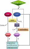
Tumour growth and metastasis: Role of NPP2-catalyzed reactions. In the extracellular milieu the tumour-motility stimulating factor NPP2 generates LPA from LPC. LPA promotes cell proliferation, migration and angiogenesis. In a coupled reaction with 5′-nucleotidase, NPP2 has the potential to hydrolyze ATP, which is cytotoxic for tumours, to adenosine, a tumour-growth promoting factor and stimulator of angiogenesis. The expression of both NPP2 and 5′-nucleotidase is increased by the Wnt/β-catenin pathway, which is activated in many cancers.

Similar articles

Cited by

References

    1. {'text': '', 'ref_index': 1, 'ids': [{'type': 'DOI', 'value': '10.1055/s-2005-869519', 'is_inner': False, 'url': 'https://doi.org/10.1055/s-2005-869519'}, {'type': 'PubMed', 'value': '15852217', 'is_inner': True, 'url': 'https://pubmed.ncbi.nlm.nih.gov/15852217/'}]}
    2. Boeynaems JM, Communi D, Gonzalez NS, Robaye B. Overview of the P2 receptors. Semin Thromb Hemost 2005; 31: 139–49. - PubMed
    1. {'text': '', 'ref_index': 1, 'ids': [{'type': 'DOI', 'value': '10.1161/hq0302.105360', 'is_inner': False, 'url': 'https://doi.org/10.1161/hq0302.105360'}, {'type': 'PubMed', 'value': '11884276', 'is_inner': True, 'url': 'https://pubmed.ncbi.nlm.nih.gov/11884276/'}]}
    2. Burnstock G. Purinergic signaling and vascular cell proliferation and death. Arterioscler Thromb Vasc Biol 2002; 22: 364–73. - PubMed
    1. {'text': '', 'ref_index': 1, 'ids': [{'type': 'DOI', 'value': '10.1007/s102380200006', 'is_inner': False, 'url': 'https://doi.org/10.1007/s102380200006'}, {'type': 'PMC', 'value': 'PMC4953172', 'is_inner': False, 'url': 'https://pmc.ncbi.nlm.nih.gov/articles/PMC4953172/'}, {'type': 'PubMed', 'value': '11871639', 'is_inner': True, 'url': 'https://pubmed.ncbi.nlm.nih.gov/11871639/'}]}
    2. Burnstock G. Potential therapeutic targets in the rapidly expanding field of purinergic signaling. Clin Med 2002; 2: 45–53. - PMC - PubMed
    1. {'text': '', 'ref_index': 1, 'ids': [{'type': 'DOI', 'value': '10.1189/jlb.1102561', 'is_inner': False, 'url': 'https://doi.org/10.1189/jlb.1102561'}, {'type': 'PubMed', 'value': '12660218', 'is_inner': True, 'url': 'https://pubmed.ncbi.nlm.nih.gov/12660218/'}]}
    2. Sak K, Boeynaems JM, Everaus H. Involvement of P2Y receptors in the differentiation of haematopoietic cells. J Leukoc Biol 2003; 73: 442–7. - PubMed
    1. {'text': '', 'ref_index': 1, 'ids': [{'type': 'DOI', 'value': '10.1016/j.brainresrev.2004.12.005', 'is_inner': False, 'url': 'https://doi.org/10.1016/j.brainresrev.2004.12.005'}, {'type': 'PubMed', 'value': '15850654', 'is_inner': True, 'url': 'https://pubmed.ncbi.nlm.nih.gov/15850654/'}]}
    2. Agresti C, Meomartini ME, Amadio S et al. ATP regulates oligodendrocyte progenitor migration, proliferation, and differentiation: Involvement of metabotropic P2 receptors. Brain Res Rev 2005; 48: 157–65. - PubMed

LinkOut - more resources