. 2006 Jun;2(2):409-30.
doi: 10.1007/s11302-006-9003-5. Epub 2006 May 30.

The E-NTPDase family of ectonucleotidases: Structure function relationships and pathophysiological significance

Affiliations

The E-NTPDase family of ectonucleotidases: Structure function relationships and pathophysiological significance

Simon C Robson et al. Purinergic Signal. 2006 Jun.

Abstract

Ectonucleotidases are ectoenzymes that hydrolyze extracellular nucleotides to the respective nucleosides. Within the past decade, ectonucleotidases belonging to several enzyme families have been discovered, cloned and characterized. In this article, we specifically address the cell surface-located members of the ecto-nucleoside triphosphate diphosphohydrolase (E-NTPDase/CD39) family (NTPDase1,2,3, and 8). The molecular identification of individual NTPDase subtypes, genetic engineering, mutational analyses, and the generation of subtype-specific antibodies have resulted in considerable insights into enzyme structure and function. These advances also allow definition of physiological and patho-physiological implications of NTPDases in a considerable variety of tissues. Biological actions of NTPDases are a consequence (at least in part) of the regulated phosphohydrolytic activity on extracellular nucleotides and consequent effects on P2-receptor signaling. It further appears that the spatial and temporal expression of NTPDases by various cell types within the vasculature, the nervous tissues and other tissues impacts on several patho-physiological processes. Examples include acute effects on cellular metabolism, adhesion, activation and migration with other protracted impacts upon developmental responses, inclusive of cellular proliferation, differentiation and apoptosis, as seen with atherosclerosis, degenerative neurological diseases and immune rejection of transplanted organs and cells. Future clinical applications are expected to involve the development of new therapeutic strategies for transplantation and various inflammatory cardiovascular, gastrointestinal and neurological diseases.


Figures

Fig. 1
Fig. 1
Hypothetical phylogenetic tree derived for 22 selected members of the E-NTPDase family (NTPDase1 to NTPDase8) from rat (r), human (h) and mouse (m), following alignment of amino acid sequences. The length of the lines indicates the differences between amino acid sequences. The graph depicts a clear separation between surface-located (top) and intracellular (bottom) NTPDases. In addition, the major substrate preferences of individual subtypes and the predicted membrane topography for each group of enzymes is given (one or two transmembrane domains, indicated by barrels). Modified from [59].
Fig. 2
Fig. 2
Cell surface-located catabolism of extracellular nucleotides and potential activation of receptors for nucleotides (P2 receptors) and adenosine (P1 receptors). The figure depicts the principal catalytic properties of members of the E-NTPDase family and of ecto-5′-nucleotidase. NTPDases sequentially convert ATP to ADP + Pi and ADP to AMP + Pi. NTPDase1 is distinct among these enzymes as it dephosphorylates ATP directly to AMP without the release of significant amounts of ADP. Hydrolysis of the nucleoside monophosphate to the nucleoside is catalyzed by ecto-5′-nucleotidase. NTPDases, NPPs and alkaline phosphatase sometimes co-exist and it seems likely that they can act in concert to metabolize extracellular nucleotides. ATP can activate both P2X receptors and subtypes P2Y receptors whereas UTP activates subtypes of P2Y receptors only. After degradation, ADP or UDP may activate additional subtypes of P2Y receptors. The adenosine formed can potentially act on four different types of P1 receptors and is either deaminated to inosine or directly recycled via nucleoside transporters. Bottom: Profiles of nucleotide hydrolysis and substrate formation by plasma membrane-located NTPDases. The figure compares catalytic properties of human and murine NTPDase1,2,3 and 8, following expression in COS-7 cells. The principal catalytic properties of the respective human and murine enzymes are similar. ATP (•), ADP (▪), AMP (≆). Modified from [57].
Fig. 3
Fig. 3
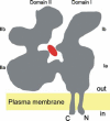
Hypothetical membrane topology of a surface-located NTPDase with two transmembrane domains. A comparison of the conserved secondary structure reveals duplicate conservation of two major domains related to subdomains Ia and IIa of actin, and other members of the actin/HSP70/sugar kinase superfamily [59]. In contrast to the other members of the superfamily, surface-located NTPDases are anchored to the plasma membrane by terminal hydrophobic domains. The figure takes into account the close distance of the N-and C-terminus of actin at domain I and the binding of ATP (red) in the cleft between domains I and II [80]. These two domains are expected to undergo conformational changes involving movement relative to each other.
Fig. 4
Fig. 4
Angiogenesis with expression of NTPDase1 in the vasculature of syngeneic islet transplants. Mouse islets were prepared from wild type and Entpd1 null mice, as described by T. Maki et al. and transplanted under the renal capsule [261]. Islets were harvested at four weeks (n = 4 per group) and stained for NTPDase1 immunoactivity and other markers of EC. Substantially diminished levels of CD31 staining vascular elements were also present in null to Entpd1 null grafts, indicating a defect in new vessel growth (not depicted here). A) Wild type to wild type showing grafted islet vasculature staining for NTPDase1 with adjacent normal renal vascular pattern. B) Wild type to null mouse showing intrinsic vasculature of islet has persisted within the graft and even entered the NTPDase1 null renal parenchyma. C) Null to wild type grafts showing infiltrating macrophages and NTPDase1 positive endothelium migrating from recipient (confirmed by other stains; not shown).
Fig. 5
Fig. 5
Detail of arrangement of neuronal stem cells and neuroblasts at the lateral lining of the mouse subventricular zone (SVZ) (triple labeling). A) DAPI staining of all nuclei. Arrow heads mark endymal lining. B) Stem cells (type B cells) immunopositive for NTPDase2 form tube-like sheeths around clusters of migrating immature neurons (type A cells) that immunostain for the microtubule-associated protein doublecortin (DCX) (C). The spaces covered by type A cells remain dark in (B) and are indicated with stars. D) Merge of B) and C). E) Merge of A), B) and C). Bar = 10 µm. (by courtesy of David Langer, Frankfurt am Main).

Similar articles

Cited by

References

    1. {'text': '', 'ref_index': 1, 'ids': [{'type': 'PubMed', 'value': '2651791', 'is_inner': True, 'url': 'https://pubmed.ncbi.nlm.nih.gov/2651791/'}]}
    2. Luthje J (1989) Origin, metabolism and function of extracellular adenine nucleotides in the blood. Klin Wochenschr 67:317–327 - PubMed
    1. {'text': '', 'ref_index': 1, 'ids': [{'type': 'PubMed', 'value': '15548415', 'is_inner': True, 'url': 'https://pubmed.ncbi.nlm.nih.gov/15548415/'}]}
    2. Burnstock G, Knight GE (2004) Cellular distribution and functions of P2 receptor subtypes in different systems. Int Rev Cytol 240:31–304 - PubMed
    1. None
    2. Lazarowski ER (2003) Molecular and biological properties of P2Y receptors. In: Schwiebert EM (ed) Extracellular Nucleotides and Nucleosides: Release, Receptors, and Physiological and Pathophysiological Effects. Academic, Amsterdam, Boston, pp 59–96
    1. {'text': '', 'ref_index': 1, 'ids': [{'type': 'PubMed', 'value': '9090307', 'is_inner': True, 'url': 'https://pubmed.ncbi.nlm.nih.gov/9090307/'}]}
    2. Harden TK, Lazarowski ER, Boucher RC (1997) Release, metabolism and interconversion of adenine and uridine nucleotides: Implications for G protein-coupled P2 receptor agonist selectivity. Trends Pharmacol Sci 18:43–46 - PubMed
    1. None
    2. Weisman GA, Erb L, Garrad RC et al (1998) P2Y nucleotide receptors in the immune system: Signaling by a P2Y2 receptor in U937 monocytes. Drug Dev Res 45:222–228