

Review
doi: 10.1002/bies.20756.
Visualizing new dimensions in Drosophila myoblast fusion
Affiliations
- PMID: 18404690
- PMCID: PMC2664634
- DOI: 10.1002/bies.20756
Item in Clipboard
Review
Visualizing new dimensions in Drosophila myoblast fusion
Bioessays.
2008 May.
Abstract
Over several years, genetic studies in the model system, Drosophila melanogastor, have uncovered genes that when mutated, lead to a block in myoblast fusion. Analyses of these gene products have suggested that Arp2/3-mediated regulation of the actin cytoskeleton is crucial to myoblast fusion in the fly. Recent advances in imaging in Drosophila embryos, both in fixed and live preparations, have led to a new appreciation of both the three-dimensional organization of the somatic mesoderm and the cell biology underlying myoblast fusion.
(c) 2008 Wiley Periodicals, Inc.
Figures
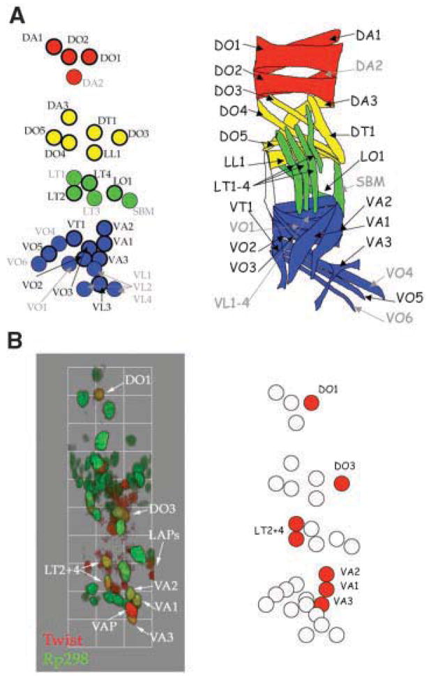
Drosophila founder cell (FC) and body wall muscle maps. A: The FC map shows the relative position of all 30 FCs prior to myoblast fusion based on three-dimensional reconstructions of single mesodermal hemisegments (left panel). The final positions of the muscles seeded by these FCs are shown in the right panel. FCs are located in four groups: dorsal (red), dorsal–lateral (yellow), lateral (green) and ventral (blue). FCs identified using the expression of FC identity markers are outlined in black, while those identified based on position are outlined in grey. DO, dorsal oblique; DA, dorsal acute; DT, dorsal transverse, LL, lateral longitudianal; LT, lateral transverse; LO, lateral oblique; SBM, segment border muscle; VT, ventral transverse; VO, ventral oblique; VA, ventral acute; VL, ventral longitudinal. B: The mesodermal transcription factor, Twist, is expressed in a subset of FCs prior to fusion. The left panel shows a three-dimensional reconstruction of a single mesodermal hemisegment at stage 13 and shows the colocalization of Twist (red) with the FC marker rp298–lacZ (green) in the FCs that will form the VA1–VA3, LT2, LT4, DO3 and DO1 muscles. A schematic of these data is shown in the panel on the right.

Cellular behaviors observed during Drosophila myoblast fusion. A: Behaviors observed by fluorescent light microscopy.(4,5,21,23,24,32) Searching: Founder cells (FCs) extend several actin-based protrusions in all directions prior to fusing. Fusion-competent myoblasts (FCMs), however, appear to extend only one actin based protrusion directionally towards an FC or myotube. Migration: FCMs assume a teardrop shape as they migrate towards an FC or myotube. An F-actin-based protrusion (red lines) extends in the direction of migration. This protrusion is broader than that observed during searching behavior. Actin focus: A spherical F-actin structure (red circle) forms at the site of adhesion between an FC or myotube and an FCM. The focus marks the subsequent site of myoblast fusion and colocalizes with several important fusion regulators. Membrane breakdown and cell mixing: Following dissolution of the actin focus, the membrane between the cells breaks down, allowing cytoplasmic and nuclear transfer and the formation of a cell with an additional nucleus. The newly fused nucleus from the FCM becomes transcriptionally “entrained” to the identity of the FC/myotube. B: Behaviors observed by transmission electron microscopy.(27) Box represents close up on the site of cell–cell adhesion, prior to fusion. Prefusion complex Electron-dense vesicles symmetrically appose across the membranes of the two cells. Electron-dense plaques: Electron-dense material presumably forms from the contents of the prefusion complex, and is also symmetrically distributed across the membranes of the two cells. Fusion pores: Gaps form as the membranes vesiculate, allowing cytoplasmic transfer and cell mixing.
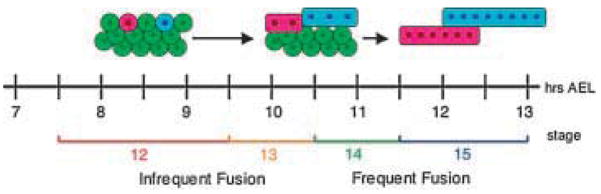
Model of Drosophila myoblast fusion. Myoblast fusion occurs between stages 12 and 15 (7.5–13 hours AEL) in two phases. At early stage 12, prior to myoblast fusion, mesodermal cells are arranged in multiple layers. The FCs (red and blue) are located externally, while the FCMs (green) are located both externally and internally. During the first phase of fusion (stage 12–13, 7.5–10.5 hours AEL), infrequent fusion events occur between the FCs and the external FCMs. The majority of fusion events occur in the second phase of fusion (stage 14–15, 10.5–13 hours AEL) as the internal FCMs dissociate from one another and migrate externally.
References
-
- Patel K, Christ B, Stockdale FE. Control of muscle size during embryonic, fetal, and adult life. Results Probl Cell Differ. 2002;38:163–186. - PubMed
-
- Abmayr SM, Balagopalan L, Galletta BJ, Hong SJ. Cell and molecular biology of myoblast fusion. Int Rev Cytol. 2003;225:33–89. - PubMed
-
- Horsley V, Pavlath GK. Forming a multinucleated cell: molecules that regulate myoblast fusion. Cells Tissues Organs. 2004;176:67–78. - PubMed
-
- Bate M. The embryonic development of larval muscles in Drosophila. Development. 1990;110:791–804. - PubMed
-
- Baylies MK, Bate M, Ruiz Gomez M. Myogenesis: a view from Drosophila. Cell. 1998;93:921–927. - PubMed
Publication types
MeSH terms
Substances
Grants and funding
LinkOut - more resources
Full Text Sources
Molecular Biology Databases
