. 2008 Apr 11:8:40.
doi: 10.1186/1471-213X-8-40.

The PI3K pathway regulates endochondral bone growth through control of hypertrophic chondrocyte differentiation

Affiliations

The PI3K pathway regulates endochondral bone growth through control of hypertrophic chondrocyte differentiation

Veronica Ulici et al. BMC Dev Biol. .

Abstract

Background: The majority of our bones develop through the process of endochondral ossification that involves chondrocyte proliferation and hypertrophic differentiation in the cartilage growth plate. A large number of growth factors and hormones have been implicated in the regulation of growth plate biology, however, less is known about the intracellular signaling pathways involved. PI3K/Akt has been identified as a major regulator of cellular proliferation, differentiation and death in multiple cell types.

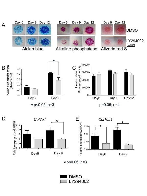
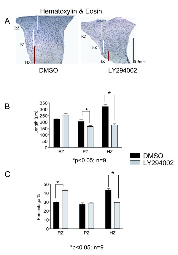
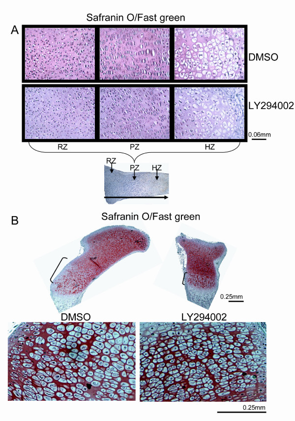
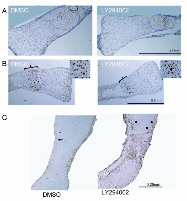
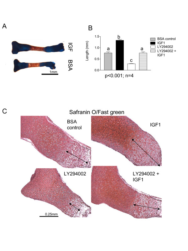
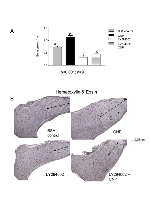
Results and discussion: Employing an organ culture system of embryonic mouse tibiae and LY294002, a pharmacological inhibitor of PI3K, we show that inhibition of the pathway results in significant growth reduction, demonstrating that PI3K is required for normal endochondral bone growth in vitro. PI3K inhibition reduces the length of the proliferating and particularly of the hypertrophic zone. Studies with organ cultures and primary chondrocytes in micromass culture show delayed hypertrophic differentiation of chondrocytes and increased apoptosis in the presence of LY294002. Surprisingly, PI3K inhibition had no strong effect on IGF1-induced bone growth, but partially blocked the anabolic effects of C-type natriuretic peptide.

Conclusion: Our data demonstrate an essential role of PI3K signaling in chondrocyte differentiation and as a consequence of this, in the endochondral bone growth process.


Figures

Figure 1
Figure 1
PI3K inhibition results in reduced chondrocyte differentiation. (A) Mesenchymal cells isolated from E11.5 mouse limb buds were cultured for 3 days and then treated with LY294002 or DMSO as vehicle control. They were stained after 6, 9 and 12 days, respectively for different chondrocyte differentiation markers: Alcian blue for glycosaminoglycans, Alkaline phosphatase activity and Alizarin red for the calcium content. The intensity of these markers is reduced in the LY294002 treated cultures. (B) After 9 and 12 days respectively the Alcian blue content of the micromasses was spectrophotometrically measured at 620 nm, after extraction with 6 M Guanidine hydrochloride. We noticed decreased absorbance levels for the LY294002 treated cultures. (C) Measurements of Hoechst fluorescence intensity (excitation/emission: 350/450 nm) showed no significant difference in the DNA content between the LY294002 and DMSO treated micromass cultures. (D, E) RNA was isolated from the micromass cultures after 6 and 9 days of culture and real time analysis was performed. The relative transcript levels for col2a1 and col10a1 were decreased in the LY294002 treated micromasses compared to the vehicle control.
Figure 2
Figure 2
Decreased growth of the LY294002 treated tibiae. (A) E 15.5 mouse tibiae, cultured for 6 days in the presence of LY294002 or DMSO, were analyzed by immunohistochemistry, at the end of the time course. Phosphorylation of Akt at Ser473 was found to be reduced under PI3K inhibition with LY294002 (10 μM). Black arrows show cells expressing phosphorylated Akt. (B) The difference between the length of the tibiae in the beginning and at the end of the time course represents the bone growth and it was found to be significantly reduced in bones treated with LY294002 (45% reduction) or PI3-K α inhibitor IV (500 nM) (35% reduction) compared to DMSO. (C) At the end of culture period the tibiae were also stained with Alcian blue/Alizarin red. We notice decreased length of both proximal (gp1) and distal (gp2) growth plates of tibiae treated with LY294002 or PI3-K α inhibitor IV. (D) Measurements of the growth plates and mineralized area (min) length confirmed the reduction of these under PI3K inhibition with LY294002. (E) When the gp1, gp2 and min were expressed as ratio of the entire bone length there is an increased ratio of the mineralized area in the LY294002 treated tibiae.
Figure 3
Figure 3
Reduced length of the proliferative and hypertrophic zones under PI3K inhibition. (A) E15.5 tibiae treated for 6 days with LY294002 or DMSO were fixed, paraffin embedded and sectioned. Hematoxylin & eosin (H&E) staining shows decreased length of hypertrophic (HZ) and proliferative zones (PZ) and increased resting zone (RZ) length in the LY294002 treated bones. (B) Measurements of three zones of the growth plate confirmed the reduction in length for the HZ and PZ in the LY294002 treated tibiae, but the RZ length zone was not found to be significantly increased. (C) The HZ length expressed as ratio from the entire growth plate length was found to be significantly decreases and the RZ significantly increased in the case of LY294002 treatment. The PZ ratio was found to be similar between the treatments.
Figure 4
Figure 4
PI3K inhibition affects chondrocyte morphology. (A) E15.5 tibiae, cultured for 6 days in the presence of LY294002 or DMSO were stained with Safranin O for proteoglycan content. The chondrocyte morphology was analyzed by comparing the chondrocyte size in the three zones of the growth plate. We noticed a reduction of hypertrophic cell size in the case of hypertrophic chondrocytes. (B) E18.5 tibiae treated with LY294002 for 6 days showed the same reduction in hypertrophic zone length and cell size as the E15.5 treated tibiae.
Figure 5
Figure 5
Decreased markers of chondrocyte differentiation and increased apoptosis in the LY294002 treated tibiae. (A) E15.5 tibia organ cultures were analyzed by immunohistochemistry for collagen X expression. The hypertrophic cell area (marked by dashed blue circles), which showed positive collagen X stain, was smaller in the LY294002 treated bones. (B) Tibiae were also analyzed by immunohistochemistry for cyclin-dependent kinase inhibitor 1C (p57), a marker of cell cycle arrest in G1 and chondrocyte differentiation. There was a narrower area of p57 positive cells (showed by brackets) in the LY294002 treated bones. (C) E 15.5 tibiae treated for 6 days with LY294002 shoed an increased number of apoptotic cells in the hypertrophic and mineralized areas (black arrows) as shown by colorimetric TUNEL assay.
Figure 6
Figure 6
IGF1-induced bone growth partially requires PI3K activity. (A)Tibiae isolated from E15.5 mice were cultured for 6 days in the presence of IGF1 or 0.1% BSA in PBS, as control. IGF1 induced significant increase in bone growth. (B) Measurements of bone growth after 6 days of treatment with IGF1, control, LY294002 or IGF1 + LY294002 showed increased bone growth in the IGF1 treatment, and similar bone growth between the control and the IGF1 + LY294002 treatment. (C) E 15.5 tibiae treated for 6 days with the treatments mentioned above, were fixed, embedded, sectioned and stained with Safranin O. The length of hypertrophic zone was increased in the IGF1 treatment compared to control; decreased in the LY294002 treatment as shown before and in the case of the IGF1 + LY294002 treatment similar to the control hypertrophic zone length.
Figure 7
Figure 7
CNP-induced bone growth requires PI3K activity. (A) Tibiae isolated from E15.5 mice were cultured for 6 days in the presence of CNP, control (0.1% HCl-BSA in PBS), LY294002 or CNP + LY294002. Bone growth measurements showed increased bone growth in the CNP treatment, and no significant difference between the LY294002 and CNP+LY294002. (B) E 15.5 tibiae treated for 6 days with the treatments mentioned above, were fixed, embedded, sectioned and stained with H&E. The length of hypertrophic zone was increased in the CNP treatment compared to control, decreased in the LY294002 treatment and similar between LY294002 and CNP+LY294002

Similar articles

Cited by

References

    1. Horton WA. Skeletal development: insights from targeting the mouse genome. Lancet. 2003;362:560–9. doi: 10.1016/S0140-6736(03)14119-0. - DOI - PubMed
    1. Shum L, Coleman CM, Hatakeyama Y, Tuan RS. Morphogenesis and dysmorphogenesis of the appendicular skeleton. Birth Defects Res C Embryo Today. 2003;69:102–22. doi: 10.1002/bdrc.10012. - DOI - PubMed
    1. Kronenberg HM. Developmental regulation of the growth plate. Nature. 2003;423:332–6. doi: 10.1038/nature01657. - DOI - PubMed
    1. Wagner EF, Karsenty G. Genetic control of skeletal development. Curr Opin Genet Dev. 2001;11:527–32. doi: 10.1016/S0959-437X(00)00228-8. - DOI - PubMed
    1. Provot S, Schipani E. Molecular mechanisms of endochondral bone development. Biochem Biophys Res Commun. 2005;328:658–65. doi: 10.1016/j.bbrc.2004.11.068. - DOI - PubMed

Publication types

MeSH terms

LinkOut - more resources