Review
. 2007 Nov;40(4):287-326.
doi: 10.1017/S0033583508004654. Epub 2008 Apr 14.

Protein folding and misfolding: mechanism and principles

Affiliations
Review

Protein folding and misfolding: mechanism and principles

S Walter Englander et al. Q Rev Biophys. 2007 Nov.

Abstract

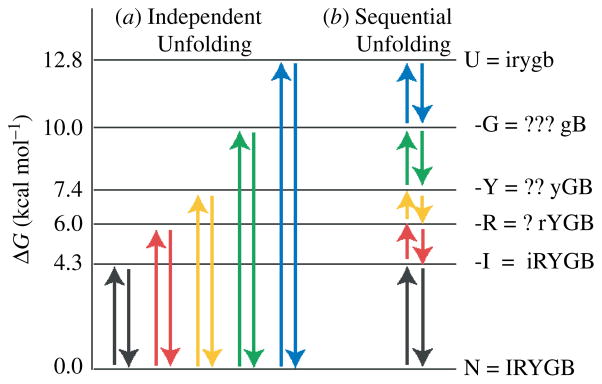
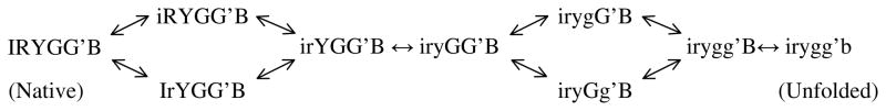
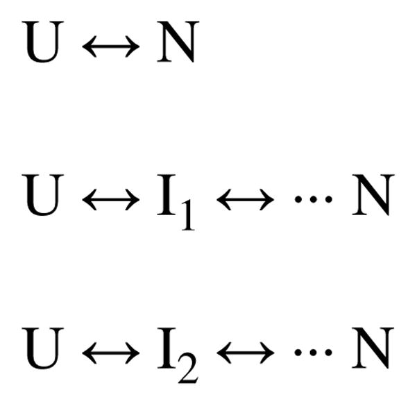
Two fundamentally different views of how proteins fold are now being debated. Do proteins fold through multiple unpredictable routes directed only by the energetically downhill nature of the folding landscape or do they fold through specific intermediates in a defined pathway that systematically puts predetermined pieces of the target native protein into place? It has now become possible to determine the structure of protein folding intermediates, evaluate their equilibrium and kinetic parameters, and establish their pathway relationships. Results obtained for many proteins have serendipitously revealed a new dimension of protein structure. Cooperative structural units of the native protein, called foldons, unfold and refold repeatedly even under native conditions. Much evidence obtained by hydrogen exchange and other methods now indicates that cooperative foldon units and not individual amino acids account for the unit steps in protein folding pathways. The formation of foldons and their ordered pathway assembly systematically puts native-like foldon building blocks into place, guided by a sequential stabilization mechanism in which prior native-like structure templates the formation of incoming foldons with complementary structure. Thus the same propensities and interactions that specify the final native state, encoded in the amino-acid sequence of every protein, determine the pathway for getting there. Experimental observations that have been interpreted differently, in terms of multiple independent pathways, appear to be due to chance misfolding errors that cause different population fractions to block at different pathway points, populate different pathway intermediates, and fold at different rates. This paper summarizes the experimental basis for these three determining principles and their consequences. Cooperative native-like foldon units and the sequential stabilization process together generate predetermined stepwise pathways. Optional misfolding errors are responsible for 3-state and heterogeneous kinetic folding.


Figures

Fig. 1
Fig. 1
Ribbon diagram of cytochrome c. Cyt c has 104 residues, three major α-helices, three major Ω-loops, and six cooperative foldon units shown in color, namely the N/C bi-helix (blue), the 60s helix and loop (green) which can be separated at low pH, the small β-sheet (yellow), and two Ω-loops (red and infrared). The two peripheral histidines that can misligate to the heme iron and block folding in a pH-sensitive way are in the green loop. Also shown are the Met80 ligand and Trp59 which serves as a FRET donor in kinetic folding experiments.
Fig. 2
Fig. 2
HX pulse-labeling results for the blocked initial intermediate in Cyt c kinetic folding. (a) The degree of labeling imposed at each measurable amide during the pulse (colored curves), analyzed by 2D NMR of the refolded native protein, can be compared with the black dashed curve expected for the case of no protection, scaled to 0·85, the fraction of the population in the intermediate state at the time of the HX pulse. The horizontal offset in the EX2 region relates to the stability against labeling. The plateau at higher pH (EX1 region) indicates the limiting kop rate. (b) Parameters of the trapped N/C bi-helical intermediate.
Fig. 3
Fig. 3
Native state hydrogen exchange. (a) Illustration of the 2-state denaturant melting curve and its analysis by the usual linear extrapolation method (Pace, 1975), showing the sharp dependence of global stability on denaturant concentration even far below the visible melting transition where NHX is measured. (b, c) The analogous measurement by NHX of transient unfolding reactions of the blue and green Cyt c foldons at low denaturant concentration. These large unfolding reactions, initially hidden by local fluctuational HX pathways (m~0), are promoted and revealed by their sharp dependence on denaturant concentration. Residues exposed to HX in each unfolding reaction join the HX isotherm and thus identify the unfolding segments. Each HX isotherm, summarized in panel (d), shows the denaturant-dependent free-energy level (relative to N) for the Cyt c partially unfolded form that first exposes each foldon unit to exchange (color coding connects to Fig. 1). Line thickening shows where HX was actually measured. The overall foldon composition of each partially unfolded form was measured in stability labeling experiments (Section 5, Fig. 5). The infrared foldon was not measured at these conditions (pDr 7, 30 °C).
Fig. 4
Fig. 4
The two extreme possibilities for foldon unfolding behavior. (a) Independent foldon unfolding. (b) Sequential foldon unfolding. Information on the foldon composition of the different partially unfolded forms revealed by the NHX results and the foldon status not yet specified by the NHX results (shown as question marks) is listed at the right.
Fig. 5
Fig. 5
Stability labeling experiments to determine partially unfolded forms composition. The modifications indicated directly alter the stability of individual foldons. The measured effect on the stability of the altered foldon (in boldface) and on the other foldons answer the question marks in Figure 4. The red and infrared foldons can unfold separately, in either order (c).
Fig. 6
Fig. 6
Kinetic folding and unfolding results for hen egg lysozyme. Kiefhaber and co-workers measured the three kinetic phases and the time-dependent populations by standard stopped-flow fluorescence methods. The data shown are globally fit by a two-pathway IUP model [extended triangular model; panels (a)–(c)] and by a PPOE model [single T model; panels (d)–(f)]. Best-fit rate constants are shown, with m values in parentheses (boldface indicates well fixed parameters). Other datasets under other conditions with additional kinetic phases were equally well fit by both models but usually with fewer fitting constants for the PPOE model.
Scheme 1
Scheme 1
Scheme 2
Scheme 2
Scheme 3
Scheme 3
Scheme 4
Scheme 4

References

    1. Anfinsen CB. Principles that govern the folding of protein chains. Science. 1973;181:223–230. - PubMed
    1. Anfinsen CB, Haber E, Sela M, White FH. Kinetics of formation of native ribonuclease during oxidation of reduced polypeptide chain. Proceedings of the National Academy of Sciences USA. 1961;47:1309–1314. - PMC - PubMed
    1. Bahar I, Wallqvist A, Covell DG, Jernigan RL. Correlation between native-state hydrogen exchange and cooperative residue fluctuations from a simple model. Biochemistry. 1998;37:1067–1075. - PubMed
    1. Bai Y, Englander JJ, Mayne L, Milne JS, Englander SW. Thermodynamic parameters from hydrogen exchange measurements. Methods in Enzymology. 1995a;259:344–356. - PubMed
    1. Bai Y, Englander SW. Future directions in folding: the multi-state nature of protein structure. Proteins: Structure, Function, Genetics. 1996;24:145–151. - PubMed

Publication types