Review
. 2008 Apr;18(2):203-17.
doi: 10.1016/j.sbi.2008.02.001. Epub 2008 Apr 9.

The structural biology of HIV assembly

Affiliations
Review

The structural biology of HIV assembly

Barbie K Ganser-Pornillos et al. Curr Opin Struct Biol. 2008 Apr.

Abstract

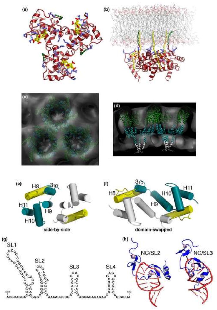
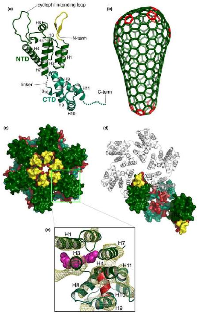
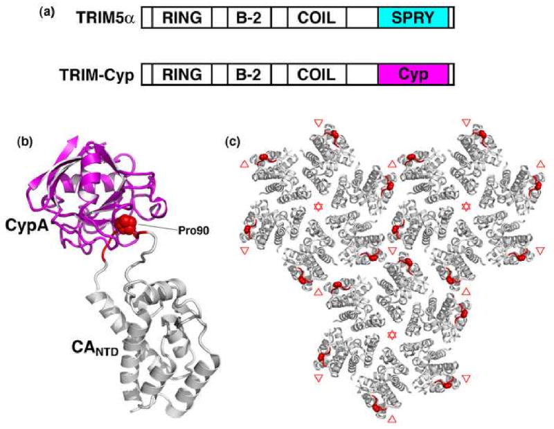
HIV assembly and replication proceed through the formation of morphologically distinct immature and mature viral capsids that are organized by the Gag polyprotein (immature) and by the fully processed CA protein (mature). The Gag polyprotein is composed of three folded polypeptides (MA, CA, and NC) and three smaller peptides (SP1, SP2, and p6) that function together to coordinate membrane binding and Gag-Gag lattice interactions in immature virions. Following budding, HIV maturation is initiated by proteolytic processing of Gag, which induces conformational changes in the CA domain and results in the assembly of the distinctive conical capsid. Retroviral capsids are organized following the principles of fullerene cones, and the hexagonal CA lattice is stabilized by three distinct interfaces. Recently identified inhibitors of viral maturation act by disrupting the final stage of Gag processing, or by inhibiting the formation of a critical intermolecular CA-CA interface in the mature capsid. Following release into a new host cell, the capsid disassembles and host cell factors can potently restrict this stage of retroviral replication. Here, we review the structures of immature and mature HIV virions, focusing on recent studies that have defined the global organization of the immature Gag lattice, identified sites likely to undergo conformational changes during maturation, revealed the molecular structure of the mature capsid lattice, demonstrated that capsid architectures are conserved, identified the first capsid assembly inhibitors, and begun to uncover the remarkable biology of the mature capsid.


Figures

Figure 1
Figure 1
(a) Summary of the HIV-1 replication cycle. (b) HIV-1 Gag polyprotein domain structure, showing the locations of MA, CANTD, CACTD, SP1, NC, SP2, and p6. (c) Structural model of the extended Gag polypeptide, derived from high-resolution structures and models of isolated domains. Unstructured and linker regions are represented by dashed lines. PR cleavage sites are indicated by the arrowheads in (b) and (c). (d, e) Schematic models of the immature (d) and mature (e) HIV-1 virions. (f, g) Central slices through cryo-EM tomograms of immature (f) and mature (g) HIV-1 particles. The spherical virions are approximately 130 nm in diameter.
Figure 2
Figure 2. Structural features of the immature Gag lattice
(a, b) Models for membrane binding by the N-terminal MA domain showing a top view (a) and a side view of the MA trimer bound to a lipid bilayer (b), with the myristoyl chain (colored in green) inserted into the inner leaflet, and basic residues (blue) interacting with acidic phospholipid headgroups, including PI(4,5)P (yellow) [25,40]. (c, d) Two views of a model based on electron cryotomography of the immature Gag lattice (gray) showing that Gag-Gag lattice interactions are mediated primarily by the CA and SP1 domains. Plausible positions of the CANTD (dark green), CACTD (blue green), and SP1 (gray) layers are shown for reference [24]. (e, f) Two crystallographically characterized HIV-1 CACTD dimers showing side-by-side (e) [68,69] and domain-swapped conformations (f) [72]. The monomers are shown in blue green and gray, and the MHR element in yellow. (g, h) Secondary structure of the HIV-1 Ψ packaging element (g), and high-resolution structures (h) of the isolated NC domain bound to the second (left) and third (right) Ψ stem loops, with NC in blue and RNA in red [85,86].
Figure 3
Figure 3. Structure of the mature HIV-1 capsid
(a) Tertiary structure of the mature, processed CA protein, with the N-terminal domain colored in dark green and the C-terminal domain in blue-green. The β-hairpin is colored yellow. The final 11 residues of CA, indicated by the dashed line are typically disordered in crystal structures. (b) Fullerene model for the conical capsid, with CA hexamers (green) and pentameric declinations (red) [89]. (c, d) Surface representation of the pseudoatomic model of the mature HIV-1 CA hexamer [102], emphasizing the CANTD hexamer (c) and CACTD dimer (d), viewed from the outer surface of the capsid. Color coding is the same as in (a), except with CA residues colored red to denote sites of significant protection from deuterium exchange in the hexagonal CA lattice [103]. Note that the exposed red patch in (d) corresponds to the CANTD-binding site on the CACTD, which is exposed when the neighboring CACTD domain is removed. (e) Top view of the CACTD-CANTD intermolecular interface. The pseudoatomic model is shown in ribbon representation, fitted to the experimental density map derived by electron cryocrystallography (gold mesh) contoured at 1.6σ [102]. Binding sites of the CA-I (red) [123] and CAP-1 (magenta) [121] maturation inhibitors are indicated.
Figure 4
Figure 4. TRIM restriction factors
(a) Domain structures of TRIM5α and TRIM-Cyp highlighting the differences in their C-terminal capsid binding domains. (b) Structure of cyclophilin A (magenta) bound to CANTD (gray) [142]. Cyclophilin-binding residues are colored red, with CA Pro90 shown explicitly. (c) Top view of the mature lattice, with three full hexamers [102]. Note that the cyclophilin-binding loops (red) surround local three-fold symmetry axes (denoted by red triangles).

References

    1. Kräusslich HG. Morphogenesis and Maturation of Retroviruses. Springer; 1996.
    1. Wills JW, Craven RC. Form, function, and use of retroviral Gag proteins. AIDS. 1991;5:639–654. - PubMed
    1. Freed EO. HIV-1 Gag proteins: diverse functions in the virus life cycle. Virology. 1998;251:1–15. - PubMed
    1. Göttlinger HG. The HIV-1 assembly machine. AIDS. 2001;15 5:S13–20. - PubMed
    1. Adamson CS, Jones IM. The molecular basis of HIV capsid assembly – five years of progress. Rev Med Virol. 2004;14:107–121. - PubMed

Publication types

Substances