Review
. 2008 Aug 1;476(1):33-42.
doi: 10.1016/j.abb.2008.03.025. Epub 2008 Mar 29.

Function, structure and regulation of the vacuolar (H+)-ATPases

Affiliations
Review

Function, structure and regulation of the vacuolar (H+)-ATPases

Kevin C Jefferies et al. Arch Biochem Biophys. .

Abstract

The vacuolar ATPases (or V-ATPases) are ATP-driven proton pumps that function to both acidify intracellular compartments and to transport protons across the plasma membrane. Intracellular V-ATPases function in such normal cellular processes as receptor-mediated endocytosis, intracellular membrane traffic, prohormone processing, protein degradation and neurotransmitter uptake, as well as in disease processes, including infection by influenza and other viruses and killing of cells by anthrax and diphtheria toxin. Plasma membrane V-ATPases are important in such physiological processes as urinary acidification, bone resorption and sperm maturation as well as in human diseases, including osteopetrosis, renal tubular acidosis and tumor metastasis. V-ATPases are large multi-subunit complexes composed of a peripheral domain (V(1)) responsible for hydrolysis of ATP and an integral domain (V(0)) that carries out proton transport. Proton transport is coupled to ATP hydrolysis by a rotary mechanism. V-ATPase activity is regulated in vivo using a number of mechanisms, including reversible dissociation of the V(1) and V(0) domains, changes in coupling efficiency of proton transport and ATP hydrolysis and changes in pump density through reversible fusion of V-ATPase containing vesicles. V-ATPases are emerging as potential drug targets in treating a number of human diseases including osteoporosis and cancer.


Figures
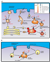
Figure 1
Figure 1. Role of intracellular V-ATPases in normal and disease processes
Panel A, role of intracellular V-ATPase in membrane trafficking, endocytosis, and secretion. Extracellular ligands are internalized by receptor mediated endocytosis and trafficked to the sorting endosome. Acidification of the endosome by the V-ATPase allows for release of the ligand and the recycling of the receptor back to the membrane. Budding of endosomal carrier vesicles and multivesicular bodies are also dependent on the acidic environment. Lysosomal proteins are synthesized in the trans Golgi network and are trafficked to the lysosome via the mannose-6-phoshpate receptor. Acidification of the late endosome allows for the release of the lysosomal proteins and recycling of the Man-6-P receptor back to the Golgi. In the lysosome acidification activates cathepsins and other degradative enzymes. The pH gradient created by the V-ATPase in secretory vesicles drives the uptake of neurotransmitters and other molecules to be secreted. The V0 domain, has also been proposed to play an important role in membrane fusion. Panel B, role of intracellular V-ATPase in the entry of envelope viruses and toxins. Envelope viruses and bacterial toxins such as diphtheria toxin enter the cell via endocytosis where they are trafficked to the sorting endosome. The low pH generated by the V-ATPase causes the viral coat to fuse with the endosomal membrane releasing the viral m-RNA into the cytoplasm. The acidic environment also induces the diphtheria toxin B chain (shown in green) to form a pore in the membrane that facilitates the entry of the A chain (shown in red) into the cytoplasm.
Figure 2
Figure 2. Function of plasma membrane V-ATPases
Panel A, function of V-ATPase in renal alpha-intercalated cells. To increase secretion of acid into the late distal tubule and collecting duct of the kidney, vesicles containing a high density of V-ATPases fuse with the apical membrane. These cells also absorb CO2, and release HCO3 into plasma through a Cl /HCO3 exchanger, preventing the cytoplasm from becoming too alkaline. Panel B, during bone resorption V-ATPase are targeted to the membranes of osteoclasts that are in contact with bone, acidifying the extracellular matrix and activating degradative enzymes. Panel C, in epididymal clear cells, V-ATPases are targeted to the luminal membrane, providing the decreased pH of the lumen required for sperm maturation. Panel D, in neutrophils and in macrophages, plasma membrane V-ATPases are involved in pH homeostasis. Panel E, in insect midgut cells, V-ATPases are used to drive a H+/2K+ antiporter resulting in the net secretion of K+ into the gut. Panel F, some invasive tumor cells target V-ATPase to the plasma membrane where they create an acidic extracellular environment that aids invasion, likely through activation of cathepsins that breakdown matrix proteins.
Figure 3
Figure 3. Structural and mechanistic features of the V-ATPase
Panel A, Schematic representation of the Saccharomyces cervisiae V-ATPase. Subunit placement and orientation are based on a combination of homology to the F-ATPase, electron microscopy studies and cysteine -mediated cross-linking studies. ATP hydrolysis by the V1 domain drives the rotation of a rotor composed of the D, F, d, and proteolipid subunits (c, c’, and c”). Rotation of the proteolipid ring past the a subunit drives the movement of protons from the cytoplasmic to the luminal side of the membrane. Two peripheral stalks, the EGC and EGH subcomplexes, hold the A3B3 hexamer stationary with respect to a. Panel B, Schematic representation of the helical arrangement in the proteolipid ring showing the unique orientation of the proteolipid subunits embedded in the membrane. The view is from the cytoplasmic side of the membrane. Panel C, Mechanism of proton translocation in the V0 domain. Only the membrane integral C-terminal domain of a (yellow) and the proteolipid ring (blue) are shown. The unprotonated form of the proteolipid’s essential glutamate residue enters the incoming hemi-channel on a. A proton from the cytoplasmic side of the membrane (the top in this diagram) enters the channel and protonates the exposed glutamic acid residue. As the proteolipid ring rotates, the now neutral glutamic acid is exposed to the hydrophobic lipid bi-layer. As the ring rotates, the glutamic acid approaches the a subunit again, and interacts with a second hemi channel on the luminal side of the membrane. Residue Arg-735 on a (shown in green) interacts with the glutamic acid, changing its pKa and promoting the deprotonation and release of the proton into the luminal side of the membrane through the hemichannel. The net result is the rotation driven pumping of protons from the cytoplasmic to the luminal side of the membrane.
Figure 4
Figure 4. Regulation of V-ATPase activity
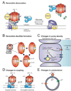
Panel A, Glucose-dependent reversible dissociation. Upon glucose depletion the V-ATPase dissociates into a soluble V1 minus subunit C, a V0, and a soluble subunit C. In their free form, neither the V1, nor the V0 has any ATPase or proton permeability, respectively. Assembly of V1 back onto V0 is mediated by the RAVE complex and aldolase, and dissociation requires intact microtubles, suggesting that dissociation and reassembly may be regulated independently. Panel B, reversible disulfide bond formation between subunit A residues Cys-254 in the catalytic site and Cys-532 locks the catalytic site into a conformation that is unable to undergo ATP hydrolysis. Panel C, changes in pump density in epididymal clear cells is regulated by HCO3 . When the luminal pH is raised, there is an incease in HCO3 , which is transported into the clear cell and activates an HCO3 sensistive adenylyl cyclase. The resulting increase in cAMP results in an increased rate of exocytosis (or decreased rate of endocytosis) of vesicles containing a high density of V-ATPases, thus increasing the density of V-ATPases at the apical membrane. While the mechanism of regulation is different, the process of vesicular fusion to deliver V-ATPase to the plasma membrane occurs in many cell types, including those shown in Figure 2. Panel D, changes in the coupling efficiency of the pump. Panel E, changes in vesicle pH though changes in Cl or H+ conductance through distinct channels.

References

    1. Forgac M. Nat. Rev. Mol. Cell. Biol. 2007;8:917–929. - PubMed
    1. Kane PM. Microbiol. Mol. Biol. Rev. 2006;70:177–191. - PMC - PubMed
    1. Nishi T, Forgac M. Nat. Rev. Mol. Cell. Biol. 2002;3:94–103. - PubMed
    1. Kakinuma Y, Ohsumi Y, Anraku Y. J. Biol. Chem. 1981;256:10859–10863. - PubMed
    1. Moriyama Y, Maeda M, Futai M. J. Exp. Biol. 1992;172:171–178. - PubMed

Publication types

MeSH terms

Substances

LinkOut - more resources