Comparative Study
. 2008 Jun;211(2):469-79.
doi: 10.1016/j.expneurol.2008.02.011. Epub 2008 Mar 2.

Transforming growth factor-beta induces cellular injury in experimental diabetic neuropathy

Affiliations
Comparative Study

Transforming growth factor-beta induces cellular injury in experimental diabetic neuropathy

Muragundla Anjaneyulu et al. Exp Neurol. 2008 Jun.

Abstract

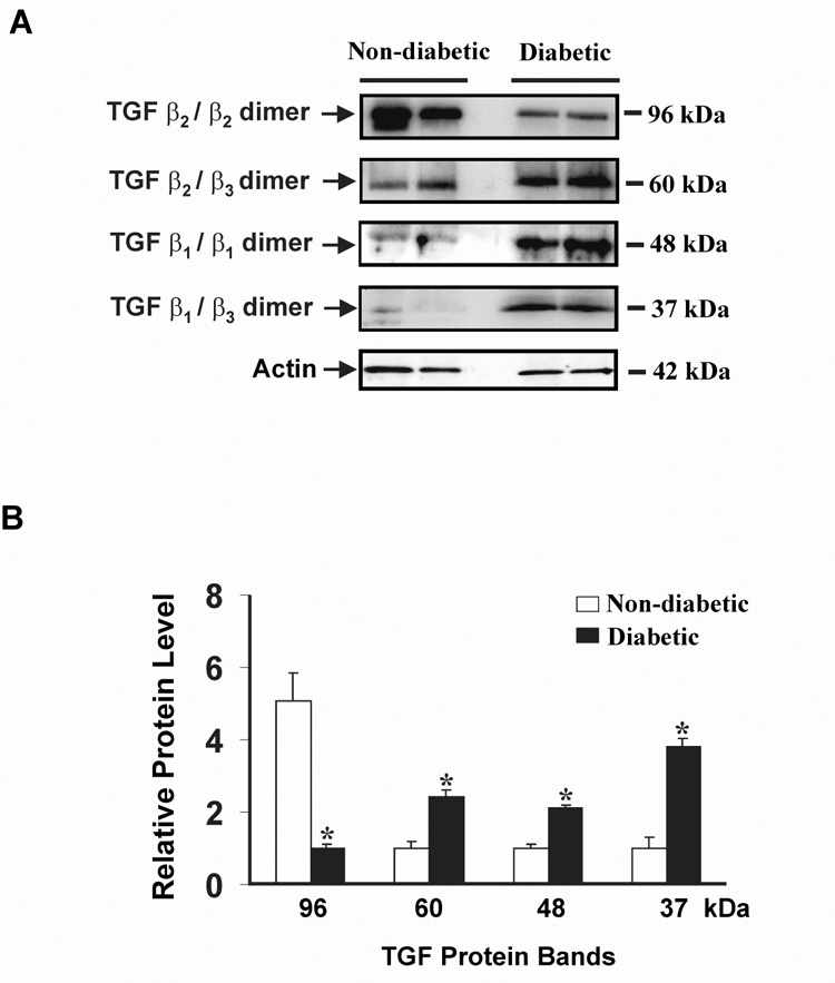
The mechanism/s leading to diabetic neuropathy are complex. Transforming growth factor-beta1 (TGF-beta1) has been associated with diabetic nephropathy and retinopathy but not neuropathy. In this study, changes in TGF-beta isoforms were examined in vivo and in vitro. Two groups of animals, streptozotocin diabetic with neuropathy and non-diabetic controls were examined at 4 weeks (n=10/group) and 12 weeks (n=8/group). In diabetic DRG using quantitative real-time PCR (QRT-PCR), TGF-beta1 and TGF-beta2 mRNA, but not TGF-beta3, was increased at 4 and 12 weeks. In sciatic nerve TGF-beta3 mRNA was primarily increased. Immunohistochemistry (DRG) and immunoblotting (sciatic nerve) showed similar differential protein expression. In sciatic nerve TGF-beta formed homo- and hetero-dimers, of which beta(2)/beta(3), beta(1)/beta(1), and beta(1)/beta(3) were significantly increased, while that of the TGF-beta(2)/beta(2) homodimer was decreased, in diabetic compared to non-diabetic rats. In vitro, pretreatment of embryonic DRG with TGF-beta neutralizing antibody prevents the increase in total TGF-beta protein observed with high glucose using immunoblotting. In high glucose conditions, combination with TGF-beta2>beta1 increases the percent of cleaved caspase-3 compared to high glucose alone and TGF-beta neutralizing antibody inhibits this increase. Furthermore, consistent with the findings in diabetic DRG and nerve, TGF-beta isoforms applied directly in vitro reduce neurite outgrowth, and this effect is partially reversed by TGF-beta neutralizing antibody. These findings implicate upregulation of TGF-beta in experimental diabetic peripheral neuropathy and indicate a novel mechanism of cellular injury related to elevated glucose levels. In combination, these findings indicate a potential new target for treatment of diabetic peripheral neuropathy.


Figures

Fig. 1
Fig. 1. Behavioral assessment of tactile allodynia in 4 week non-diabetic control and STZ-induced diabetic rats using electronic Von-Frey testing
Paw mechanical withdrawal threshold (g) was significantly decreased in diabetic rats. Data is expressed as mean ± SEM. **P<0.001 as compared with non-diabetic control.
Fig. 2
Fig. 2. Paw skin biopsy for nerve fibers
Images show sections immunostained with anti-PGP 9.5 antibody in 50 µm cryosections from the hind limb. The arrow indicates intraepidermal nerve fibers; arrow heads indicate dermal nerve fibers (scale bar 30 µm). There is a marked reduction in IENFD in the diabetic rats (B) as compared with the non-diabetic control (A). Lower panel (C) indicates the measurement of IENFD both in non-diabetic and diabetic animals. **P<0.001 as compared to non-diabetic control animals.
Fig. 3
Fig. 3. Expression of different isoforms of TGF-β in 4 & 12 week non-diabetic control and STZ-induced diabetic rats in DRG (A – 4 weeks, B – 12 weeks) and sciatic nerve (C – 4 weeks, D – 12 weeks)
QRT-PCR was performed using specific primers as described in experimental procedures. TGF-β1 and TGF-β2, but not TGF-β3 was increased in DRG. In sciatic nerve, only TGF-β3 is increased. Data is expressed as mean ± SEM. *P<0.05 & **P<0.001 as compared with non-diabetic control.
Fig. 4
Fig. 4. Immunohistochemistry of 4 weeks non-diabetic control and diabetic rat DRG tissue
(A) Light microscopic images: a, c, e; immunofluoresence (FITC staining) images: b, d, f. TGF-β2 was significantly increased in diabetic DRG (d) compared to non-diabetic control (b) There was no immunofluorescence using pretreatment with pan- TGF-β neutralizing antibody on the section as an internal control (f). (B) Intensity measurement: TGF-β2 staining was significantly increased in diabetic DRG (**P<0.001).
Fig. 5
Fig. 5. Western blot analysis of TGF-β isoforms in 4 week non-diabetic and diabetic rat sciatic nerve
(A) Lane 1 & 2 represents non-diabetic protein bands; lane 3 & 4 represents protein bands from diabetic rats. In this blot actin-β was used as an internal control. (B) The density of the relative protein levels were measured and expressed as a graph. Clear bars indicate the relative protein level in non-diabetic controls, black bars indicate the relative protein level in diabetic rats. Data is expressed as mean ± SEM. *P<0.05 as compared with non-diabetic rats.
Fig. 6
Fig. 6. Pretreatment with TGF-β neutralizing antibody normalizes the increase in total TGF-β antibody levels with high glucose
Western immunoblot analysis of TGF-β was performed in pooled cultured embryonic DRG neurons with high glucose ± neutralizing antibody: (A) Control DRG neurons (lane 1), high glucose treated neurons (lane 2), high glucose plus TGF-β neutralizing antibody (Lane 3), TGF-β control protein (lane 4). In this blot actin-β was used as internal control. (B) The density of the relative protein levels were measured and expressed as a graph. Data is expressed as mean ± SEM. *P<0.05 as compared with control, #P<0.05 as compared with high glucose,
Fig. 7
Fig. 7. Increased glucose levels and purified combined TGF-β induces DRG neuronal injury
Purified DRG neurons were treated for 24 hours with high glucose, and/or purified TGF-β protein and stained and counted for caspase-3 cleavage. There is an increase in the percent of caspase-3 positive neurons in DRG neurons exposed to high glucose or TGF-β in a dose dependent manner and there is an additive effect with glucose and higher doses of TGF-β. Data is expressed as mean ± SEM. ***, ###P<0.01 as compared with control, +++P<0.01 as compared with high glucose treatment.
Fig. 8
Fig. 8. TGF-β1, -β2, & -β3 increases glucose-induced neuronal injury
(A) A higher percentage of cultured DRG neurons expressed cleaved caspase-3 after incubation in high glucose + TGF-β1, -β2 & -β3 for 24 hours than treated or non-treated controls. Addition of glucose to TGF-β1, -β2 & -β3 had an additive effect on the percent of caspase-3 positive neurons. TGF-β Neutralizing antibody prevented glucose-induced caspase-3 cleavage. Data expressed as mean ± SEM. #P<0.05 as compared with non-treated control cells, *P<0.05, **P<0.01 in glucose-treated neurons as compared with the corresponding control condition. (B) DRG neurons that were exposed to high glucose for 48 hours had a higher rate of neuronal injury than non-treated control cells as indicated by TUNEL staining of nuclei for DNA degradation. TGF-β Neutralizing antibody prevented glucose-induced neuronal injury. Data is expressed as mean ± SEM. **P<0.001 in glucose-treated neurons as compared to control.
Fig. 9
Fig. 9. TGF-β1, -β2, & -β3 reduce neurite outgrowth
(A) DRG explants cultured with 10 ng/ml of purified TGF-β1, -β2, or -β3 for 48 hours had a reduced mean neurite length compared to control explants. Data expressed as mean ± SEM. (B) high glucose reduces neurite outgrowth but this effect is partially reversed by the use of the TGF-β neutralizing antibody. TGF-β neutralizing antibody alone does not affect neurite growth.

References

    1. Barna G, Sebestyen A, Chinopoulos CC, Nagy K, Mihalik R, Paku S, Kopper L. TGF beta 1 kills lymphoma cells using mitochondrial apoptotic pathway with the help of caspase-8. Anticancer Res. 2002;22:3867–3872. - PubMed
    1. Berent-Spillson A, Robinson A, Golovoy D, Slusher B, Rojas C, Russell JW. Protection against glucose-induced neuronal death by NAAG and GCP II inhibition is regulated by mGluR3. J. Neurochem. 2004;89:90–99. - PubMed
    1. Berent-Spillson A, Russell JW. Metabotropic glutamate receptor 3 protects neurons from glucose-induced oxidative injury by increasing intracellular glutathione concentration. J. Neurochem. 2007;101:342–354. - PubMed
    1. Bianchi R, Buyukakilli B, Brines M, Savino C, Cavaletti G, Oggioni N, Lauria G, Borgna M, Lombardi R, Cimen B, Comelekoglu U, Kanik A, Tataroglu C, Cerami A, Ghezzi P. Erythropoietin both protects from and reverses experimental diabetic neuropathy. Proc. Natl. Acad. Sci. U. S. A. %20. 2004;101:823–828. - PMC - PubMed
    1. Blobe GC, Schiemann WP, Lodish HF. Role of transforming growth factor beta in human disease. N. Engl. J. Med. 2000;342:1350–1358. - PubMed

Publication types

MeSH terms