Review
. 2008 Jun-Jul;97(2-3):163-79.
doi: 10.1016/j.pbiomolbio.2008.02.005. Epub 2008 Feb 13.

Tensegrity-based mechanosensing from macro to micro

Affiliations
Review

Tensegrity-based mechanosensing from macro to micro

Donald E Ingber. Prog Biophys Mol Biol. 2008 Jun-Jul.

Abstract

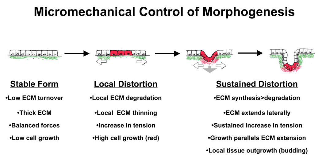
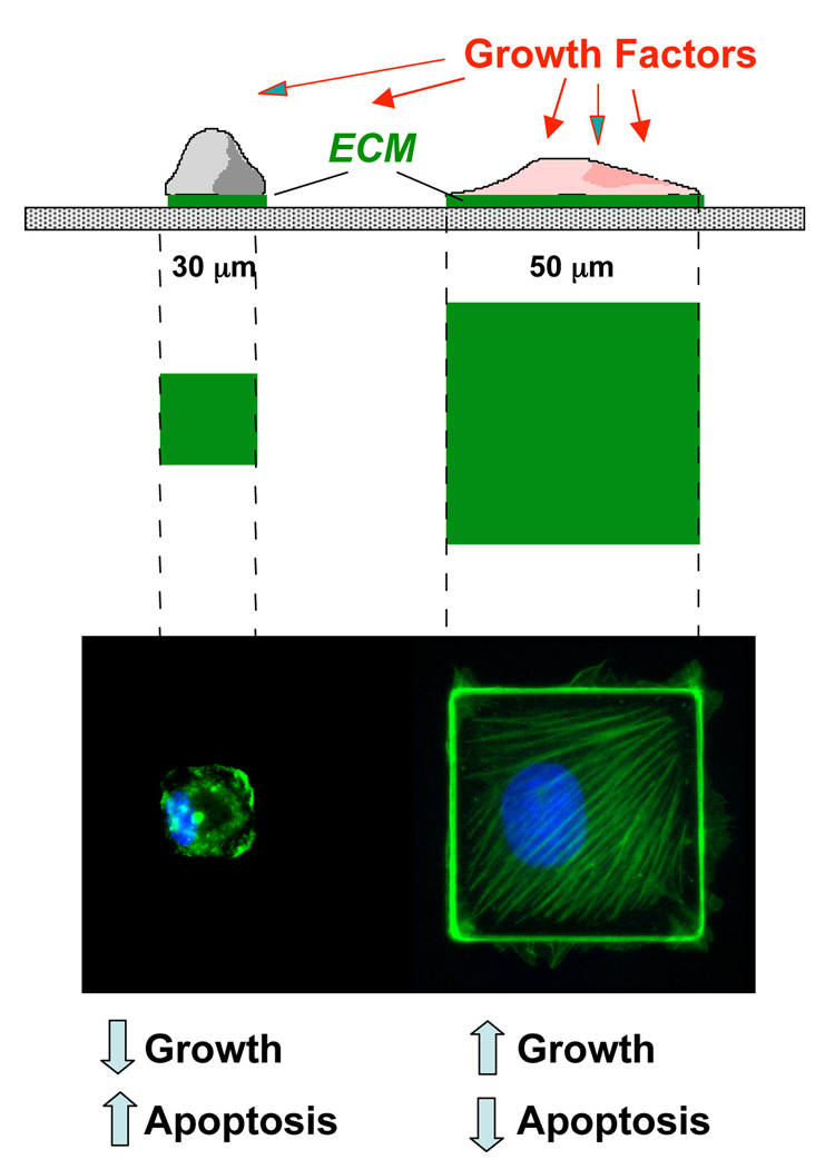
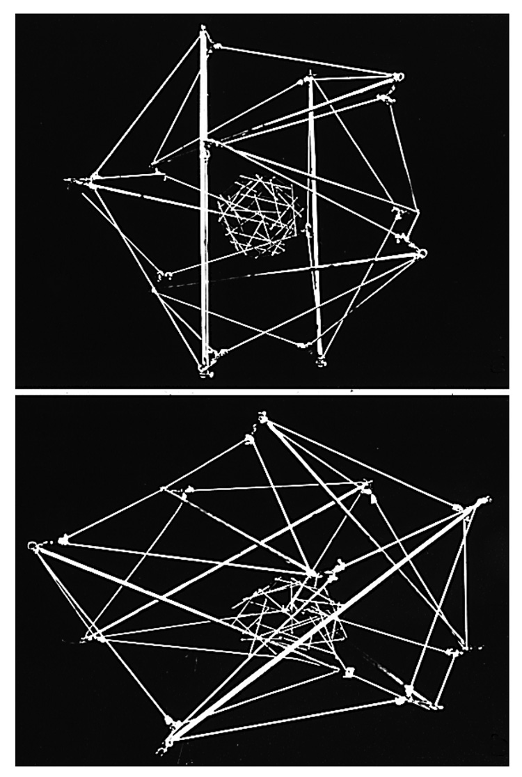
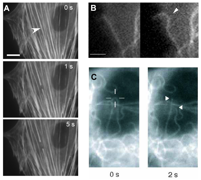
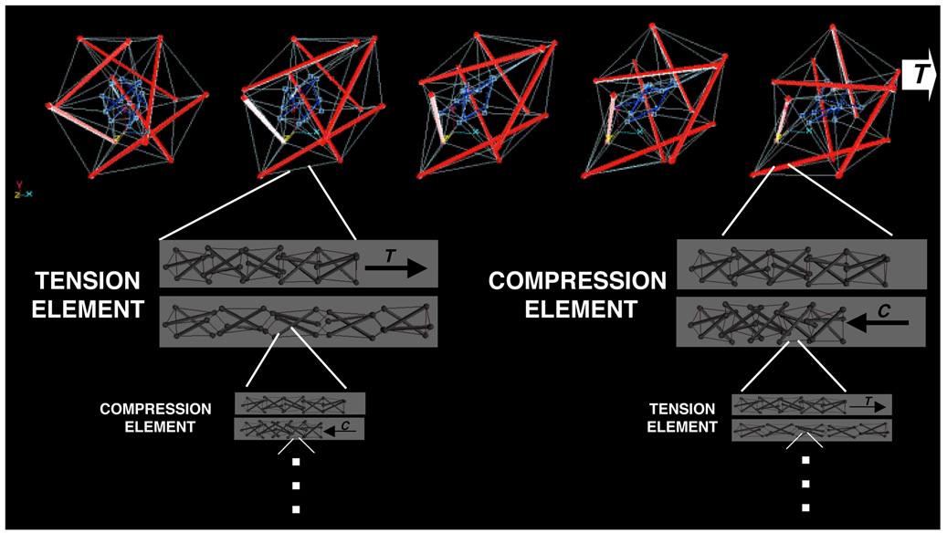
This article is a summary of a lecture on cellular mechanotransduction that was presented at a symposium on "Cardiac Mechano-Electric Feedback and Arrhythmias" that convened at Oxford, England in April 2007. Although critical mechanosensitive molecules and cellular components, such as integrins, stretch-activated ion channels, and cytoskeletal filaments, have been shown to contribute to the response by which cells convert mechanical signals into a biochemical response, little is known about how they function in the structural context of living cells, tissues and organs to produce orchestrated changes in cell behavior in response to stress. Here, studies are reviewed that suggest our bodies use structural hierarchies (systems within systems) composed of interconnected extracellular matrix and cytoskeletal networks that span from the macroscale to the nanoscale to focus stresses on specific mechanotransducer molecules. A key feature of these networks is that they are in a state of isometric tension (i.e., experience a tensile prestress), which ensures that various molecular-scale mechanochemical transduction mechanisms proceed simultaneously and produce a concerted response. These features of living architecture are the same principles that govern tensegrity (tensional integrity) architecture, and mathematical models based on tensegrity are beginning to provide new and useful descriptions of living materials, including mammalian cells. This article reviews how the use of tensegrity at multiple size scales in our bodies guides mechanical force transfer from the macro to the micro, as well as how it facilitates conversion of mechanical signals into changes in ion flux, molecular binding kinetics, signal transduction, gene transcription, cell fate switching and developmental patterning.


Figures

Fig. 1
Fig. 1. Micromechanical Control of Tissue Morphogenesis
Diagrams of a model for tension-driven tissue remodeling during normal epithelial morphogenesis. Local increases in ECM turnover result in formation of a focal defect in the basement membrane (green) that stretches and thins due to the contraction and pulling of neighboring adherent epithelium (white arrows) and underlying mesenchyme (gray arrow). Cells adherent to the basement membrane in this extending region will distort or experience changes in tension within the cytoskeleton and thus, become preferentially sensitive to growth stimuli. Cell division is accompanied by deposition of new basement membrane (red) and thus, cell mass expansion and ECM extension are tightly coupled leading to bud formation in this localized area of the developing tissue (modified from Huang and Ingber, 1999).
Fig. 2
Fig. 2. Control of Cell Shape and Function using Micropatterned Adhesive Substrates containing Micrometer-Sized ECM Islands
Top) Side view of a schematic design of a cell culture substrate containing adhesive islands of defined shape and size on the micron scale that were coated with a saturating density of fibronectin (green) and separated by intervening non-adhesive regions coated with polyethylene glycol using a self assembly-based microfabrication method. Middle) A view from above showing the same micropatterned adhesive islands. Bottom) Immunofluorescence micrographs of endothelial cells cultured on the corresponding islands shown above and stained for actin microfilaments with FITC-phalloidin (green) and DNA with DAPI (blue). Note that cells remain small and are devoid of large actin bundles on the small adhesive island, but spread and form well organized stress fibers oriented diagonally when cultured on a large island. Under these conditions, spread cells on large islands pass through the late G1 checkpoint when stimulated by growth factors, whereas endothelial cells that are restricted in their spreading never enter S phase, and instead undergo apoptosis.
Fig. 3
Fig. 3. Tensegrity Cell Model
A large tensegrity structure built as a model of a nucleated mammalian cell that was constructed from aluminum struts and thick elastic cord, with a geodesic sphere composed of wooden sticks and thin white elastic thread at its center. The cell and nucleus are interconnected by thin black elastic thread that can not be seen due to the black background. Top) Cell and nuclear shape are both round in a symmetrical cell that generates internal tension and is unanchored. Bottom) The tensegrity cell and nucleus extend in a coordinated fashion when attached to a rigid substrate, and the nucleus polarizes (moves to the base) because of tensional continuity in the structure (reprinted with permission from Ingber, 1993).
Fig. 4
Fig. 4. Mechanical Behavior of Cytoskeletal Filaments in Living Cells
A) Severing and spontaneous retraction of a single stress fiber bundle in an endothelial cell expressing EYFP-actin using a femtosecond-based nanoscissor reveals prestress in these cytoskeletal bundles (arrowhead indicates position of the laser spot; bar = 10 µm; from Kumar et al., 2006). B) Fluorescence video microscopy images of a cell expressing GFP-tubulin showing buckling of a microtubule (arrowhead) as it polymerizes and impinges end-on on the cell cortex (right vs. left; bar = 2 µm; from Wang et al., PNAS 2001). C) Fluorescence micrographs of a GFP-labeled microtubule in an endothelial cell before (left) and 2 sec after (right) it was incised with the laser nanoscissor. Note that the previously bent microtubule rapidly snaps back to a straight shape immediately after it is cut; the cross hair shows the position targeted by the laser (from Heisterkamp et al., 2005).
Fig. 5
Fig. 5. Computer models depicting multiscale structural rearrangements with a prestressed tensegrity hierarchy
Top) A two-tier hierarchical tensegrity composed of concentric large (red) and small (blue) spherical (polyhedral) strut and cable structures connected by tension elements. Note that the structure exhibits coordinated structural rearrangements of its internal elements as it extends to the right in response to tension (T) application (movies showing dynamic movements in tensegrities can be seen at: www.childrenshospital.org/research/Site2029/mainpageS2029P23sublevel24.html). Lower panels show how individual struts and cables of the structure may themselves be organized as compressed (C) and tensed (T) tensegrity mast structures at smaller and smaller size scales ad infinitum. A stress applied at the macroscale will result in global rearrangements at multiple size scales, rather than local bending or breakage, as long as tensional integrity and stabilizing prestress are maintained throughout the hierarchical network (reprinted with permission from Ingber, 2006).

References

    1. Alenghat FJ, Ingber DE. Mechanotransduction: all signals point to cytoskeleton, matrix, and integrins. Sci STKE. 2002. 2002:PE6. - PubMed
    1. Alenghat FJ, Nauli SM, Kolb R, Zhou J, Ingber DE. Global cytoskeletal control of mechanotransduction in kidney epithelial cells. Exp Cell Res. 2004;301:23–30. - PubMed
    1. Ausprunk DH, Folkman J. Migration and proliferation of endothelial cells in preformed and newly formed blood vessels during tumor angiogenesis. Microvasc Res. 1977;14:53–65. - PubMed
    1. Bernfield MR, Banerjee SD. The basal lamina in epithelial-mesenchymal interactions. In: Kefalides N, editor. Biology and Chemistry of Basement Membranes. New York: Academic Press; 1978. pp. 137–148.
    1. Brangwynne CMF, Kumar S, Geisse NA, Mahadevan L, Parker KK, Ingber DE, Weitz D. Microtubules can bear enhanced compressive loads in living cells due to lateral reinforcement. J. Cell Biol. 2006 in press. - PMC - PubMed

Publication types