. 2008 May 15;153(3):842-50.
doi: 10.1016/j.neuroscience.2008.03.003. Epub 2008 Mar 8.

Tonic and stimulus-evoked nitric oxide production in the mouse olfactory bulb

Affiliations

Tonic and stimulus-evoked nitric oxide production in the mouse olfactory bulb

G Lowe et al. Neuroscience. .

Abstract

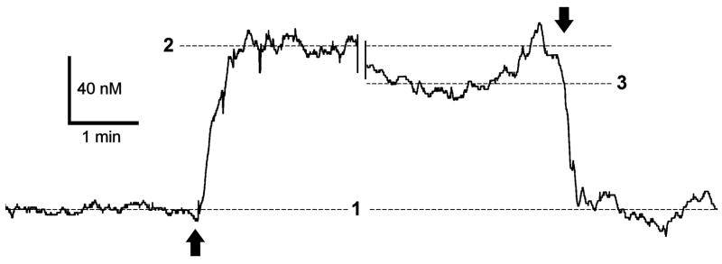
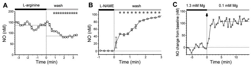
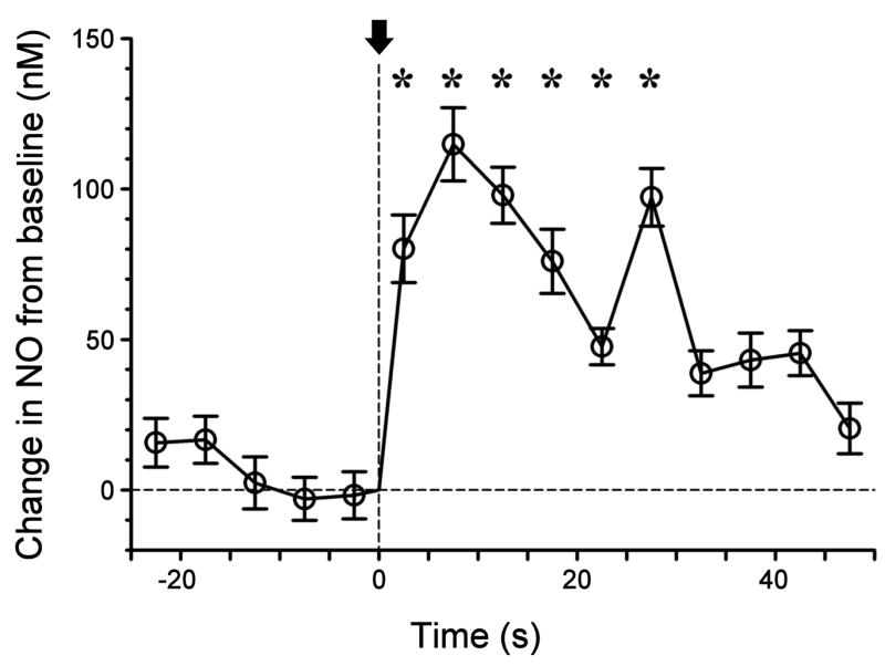
Nitric oxide (NO) has been long assumed to play a key role in mammalian olfaction. This was based largely on circumstantial evidence, i.e. prominent staining for nitric oxide synthase (NOS) and cyclic guanosine 3',5'-cyclic monophosphate (cGMP) or soluble guanylyl cyclase, an effector enzyme activated by NO, in local interneurons of the olfactory bulb. Here we employ innovative custom-fabricated NO micro-sensors to obtain the first direct, time-resolved measurements of NO signaling in the olfactory bulb. In 400 microm thick mouse olfactory bulb slices, we detected a steady average basal level of 87 nM NO in the extracellular space of mitral or granule cell layers. This NO 'tone' was sensitive to NOS substrate manipulation (200 microM L-arginine, 2 mM N(G)-nitro-L-arginine methyl ester) and Mg(2+) modulation of N-methyl-D-aspartate (NMDA) receptor conductance. Electrical stimulation of olfactory nerve fibers evoked transient (peak at 10 s) increments in NO levels 90-100 nM above baseline. In the anesthetized mouse, NO micro-sensors inserted into the granule cell layer detected NO transients averaging 55 nM in amplitude and peaking at 3.4 s after onset of a 5 s odorant stimulation. These findings suggest dual roles for NO signaling in the olfactory bulb: tonic inhibitory control of principal neurons, and regulation of circuit dynamics during odor information processing.


Figures

Figure 1
Figure 1. Nitric oxide gradients in the mouse olfactory bulb slice
A calibrated NO micro-sensor was positioned in the slice perfusion chamber with the tip initially located ~300 μm above the slice surface. The sensor was subsequently driven along its axis with a micromanipulator until the tip penetrated the slice near the mitral cell body layer to a depth of ~ 50 μm. Electrode current was sampled at 10 Hz during this procedure. After a 42 minute gap in the record during which several electrical stimulation trials were done, the NO sensor tip was retracted from the slice. The up and down arrows indicate the times of sensor penetration and retraction, respectively. Dashed lines indicate mean NO readings before (1) and a few minutes after (2) slice penetration, and a few minutes before retraction (3).
Figure 2
Figure 2. Tonic nitric oxide production in the mouse olfactory bulb slice is altered by modulating NOS and neuronal activity
A. Removal of the nNOS substrate L-arginine (at time 0) resulted in a significant decrease in NO micro-sensor signal, which settled to a new steady current. Starred values are significantly less than mean pre-wash out values (p < 0.05, Mann Whitney rank sum test). Horizontal dashed lines show mean steady state values before and after perfusion switch. B. Removal of L-NAME, a competitive inhibitor of nNOS, resulted in a significant increase in NO micro-sensor signal (zero current corresponding to zero NO level for this case, as L-NAME completely inhibited NO synthesis). Starred values are significantly higher than mean pre-wash out value (p < 0.05, Mann Whitney rank sum test). Data in A and B were acquired at 10 Hz and binned in 15 s intervals (mean values with standard deviations indicated). Horizontal dashed line shows mean steady state value before perfusion switch. Electrode calibration was 20 nM/pA. C. An NO micro-sensor was inserted into the granule cell layer of a slice (depth ~ 50 μm) and electrode current was recorded as the slice perfusion solution was switched from 1.3 mM Mg2+ to 100 μM Mg2+ (arrow, time 0) which increases excitability of neurons. Current was sampled at 20 Hz and binned in 30 s intervals bins (mean values with standard deviations indicated). Horizontal dashed lines show mean steady state values before and after perfusion switch. The time delay between valve switch and completion of solution change is 1.5 min. Electrode calibration was 24 nM/pA.
Figure 3
Figure 3. Electrical stimulation evokes nitric oxide production in the mouse olfactory bulb slice
An NO micro-sensor was inserted into the granule cell layer of a slice (depth ~ 50 μm) and the electrode current was recorded as the slice was stimulated by current pulses from concentric bipolar electrode placed in olfactory nerve layer radial to the sensor site (a 200 ms train of 20 current pulses at 100 Hz, initiated at time 0, indicated by arrow). The plot shows data sampled at 20 Hz binned in 5 s intervals (mean values with standard deviations indicated). Starred values are significantly greater than mean pre-shock values (p < 0.05, Mann Whitney rank sum test). Electrode calibration was 24 nM/pA.
Figure 4
Figure 4. Odorant-evoked nitric oxide transients in the anesthetized mouse olfactory bulb
A. Signal recorded from a calibrated NO micro-sensor inserted into the granule cell layer at a depth of 500 μm relative to the surface of the dura. The plot is the averaged response for multiple trials applying 6 of 7 odorant mixtures (Table 1). B. Signal recorded from the granule cell layer at a depth of 900 μm (averaged response for all 7 odorant mixtures in Table 1). Signal was sampled at 100 Hz and then averaged in intervals of 500 ms. Bars indicate standard error of the mean. Dashed lines indicate the period of actuation of solenoid valves controlling odorant stimulation.

References

    1. Ahern GP, Klyachko VA, Jackson MB. cGMP and S-nitrosylation: two routes for modulation of neuronal excitability by NO. Trends Neurosci. 2002;25:510–517. - PubMed
    1. Allen BW, Piantadosi CA, Coury LAJ. Electrode materials for nitric oxide detection. Nitric Oxide: Biology and Chemistry. 2000;4:75–84. - PubMed
    1. Alonso JR, Porteros A, Crespo C, Arevalo R, Brinon JG, Weruaga E, Aijon J. Chemical anatomy of the macaque monkey olfactory bulb: NADPH-diaphorase/nitric oxide synthase activity. J Comp Neurol. 1998;402:419–434. - PubMed
    1. Bohlen HG. Mechanism of increased vessel wall nitric oxide concentrations during intestinal absorption. Am J Physiol Heart Circ Physiol. 1998;275:H542–H550. - PubMed
    1. Bredt DS, Glatt CE, Hwang PM, Fotuhi M, Dawson TM, Snyder SH. Nitric oxide synthase protein and mRNA are discretely localized in neuronal populations of the mammalian CNS together with NADPH diaphorase. Neuron. 1991;7:615–624. - PubMed

Publication types