. 2008 Aug;237(8):2021-9.
doi: 10.1002/dvdy.21531.

Identification of genes involved in the ciliary trafficking of C. elegans PKD-2

Affiliations

Identification of genes involved in the ciliary trafficking of C. elegans PKD-2

Young-Kyung Bae et al. Dev Dyn. 2008 Aug.

Abstract

Ciliary membrane proteins are important extracellular sensors, and defects in their localization may have profound developmental and physiological consequences. To determine how sensory receptors localize to cilia, we performed a forward genetic screen and identified 11 mutants with defects in the ciliary localization (cil) of C. elegans PKD-2, a transient receptor potential polycystin (TRPP) channel. Class A cil mutants exhibit defects in PKD-2::GFP somatodendritic localization while Class B cil mutants abnormally accumulate PKD-2::GFP in cilia. Further characterization reveals that some genes mutated in cil mutants act in a tissue-specific manner while others are likely to play more general roles in such processes as intraflagellar transport (IFT). To this end, we identified a Class B mutation that disrupts the function of the cytoplasmic dynein light intermediate chain gene xbx-1. Identification of the remaining mutations will reveal novel molecular pathways required for ciliary receptor localization and provide further insight into mechanisms of ciliary signaling.


Figures

Figure 1
Figure 1
Identification of new genes that regulate PKD-2::GFP ciliary localization in sensory neurons. A) Schematic of a C. elegans sensory neuron expressing PKD-2::GFP. PKD-2::GFP localizes to the cell body ER and cilium (including the cilium proper and ciliary base) and small moving dendritic puncta. Arrows in the dendrite indicate that these puncta move in both anterograde and retrograde directions. B) F1 and F2 clonal screens were performed with EMS mutagenized myIs1[Ppkd-2::PKD-2::GFP; Pcc::GFP] pkd-2(sy606)IV; him-5(e1490)V hermaphrodites. F2 or F3 adult males were scored for PKD-2::GFP localization defects in head CEM and tail ray neurons. In total, 11 mutants were identified from 1032 haploid genomes. C) xbx-1 genomic region, mutations, and the XBX-1 protein which is comprised of a single dynein light intermediate chain (DLIC) domain.
Figure 2
Figure 2
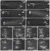
PKD-2::GFP localization in the CEM neurons of wild type (A, G), Class A (B, C, H, I, J), and Class B (D, E, F, K, L, M, N) mutants. In all panels, double arrowheads indicate the cilium proper («), arrowheads indicate the ciliary base (σ), and arrows indicate the dendrite. A-F show the distribution of PKD-2::GFP throughout the entire neuron (scale bar 10μm). Images in panels A-F are projected confocal images. Panels G-N are generated from projected epifluorescent images after 3D devolution overlayed on Normarski pictures. A) In wild-type males, PKD-2::GFP is localized in the ER of cell bodies (double-headed arrow), the cilium proper («) and the ciliary base (σ), which corresponds to the distal most dendritic ending and ciliary transition zone. B) In cil-2(my2) mutants, PKD-2::GFP is mislocalized throughout the dendrite and small GFP particles are visible outside the dendrite (near arrow). PKD-2::GFP accumulates at the ciliary base. C) my12 mutants exhibit punctate PKD-2::GFP localization along the dendrite (near arrow). PKD-2::GFP accumulates in the cilium proper and within the ciliary base. D) my14 mutants exhibit increased PKD-2::GFP in the cilium proper and the ciliary base. E) In my16 males, PKD-2::GFP localization accumulates at the ciliary base. (F) PKD-2::GFP accumulates in the ciliary base of xbx-1(my17) mutants. Panels G-N exhibit the localization of PKD-2::GFP in the ciliary region (compare with Table 2; scale bar 5μm). Panels G-N, a yellow dotted line indicates the outline of the buccal cavity. The ciliary base is located midway between the anterior and posterior ends of the cavity. G) In wild-type CEM neurons, PKD-2::GFP localizes to the ciliary base and cilium proper. H) In a my9 mutant, PKD-2::GFP accumulates at the ciliary base and in the cilium proper. A double-arrow points out PKD-2::GFP localization in the distal dendrite (underlying the buccal cavity) and more proximal dendrite. I) In a my11 mutant, PKD-2::GFP accumulates in the cilium proper. Both the cilium proper and the base exhibit morphological defects such as a hook-like projection (open arrowhead). PKD-2::GFP is visible in the dendrite (arrow). J) In a my22 mutant, PKD-2::GFP accumulates in the cilium proper. There are increased numbers of PKD-2::GFP puncta in the cilium proper, ciliary base, and dendrite. K) In a cil-5(my13) mutant, the cilium bends inward towards the buccal cavity, much like a kap-1 or klp-11 mutant (Bae et al., 2006). PKD-2::GFP puncta accumulate at the ciliary base. L) In a my14 mutant, PKD-2::GFP levels are increased in the cilium proper, which is often bent inward like cil-5(my13). The morphology of the ciliary base is also abnormal: hook-like structures are often visible (open arrowhead). M) In a my16 mutant, PKD-2::GFP accumulates in small puncta at the ciliary base. PKD-2::GFP labeling is also visible in the distal, but not proximal dendrites (double arrows). N) In an xbx-1(my17) mutant, PKD-2::GFP aggregates at the ciliary base and PKD-2::GFP localization extends into the distal (not proximal) dendrite (double arrows). When visible, the xbx-1(my17) cilium proper appears stunted.
Figure 3
Figure 3
PKD-2::GFP localization in ray neurons. A) In wild-type, PKD-2::GFP is found in the cell bodies and ciliary regions (arrowheads) at the ray tips. B, C, D) In my22, cil-5(my13) and my16 males, PKD-2::GFP similarly accumulates along the distal part of ray dendrites (two arrows). These images are projected epifluorescent images after 3D deconvolution. Scale bar 5μm.

References

    1. Anderson P. Mutagenesis. Methods Cell Biol. 1995;48:31–58. - PubMed
    1. Bae YK, Qin H, Knobel KM, Hu J, Rosenbaum JL, Barr MM. General and cell-type specific mechanisms target TRPP2/PKD-2 to cilia. Development. 2006;133:3859–3870. - PubMed
    1. Bargmann CI. Comparative chemosensation from receptors to ecology. Nature. 2006;444:295–301. - PubMed
    1. Barr MM, DeModena J, Braun D, Nguyen CQ, Hall DH, Sternberg PW. The Caenorhabditis elegans autosomal dominant polycystic kidney disease gene homologs lov-1 and pkd-2 act in the same pathway. Curr Biol. 2001;11:1341–1346. - PubMed
    1. Barr MM, Sternberg PW. A polycystic kidney-disease gene homologue required for male mating behaviour in C. elegans. Nature. 1999;401:386–389. - PubMed

Publication types

MeSH terms

LinkOut - more resources