Review
. 2008 Aug;237(8):2007-12.
doi: 10.1002/dvdy.21530.

Cholangiocyte primary cilia in liver health and disease

Affiliations
Review

Cholangiocyte primary cilia in liver health and disease

Anatoliy I Masyuk et al. Dev Dyn. 2008 Aug.

Abstract

The epithelial cells lining intrahepatic bile ducts (i.e., cholangiocytes), like many cell types in the body, have primary cilia extending from the apical plasma membrane into the bile ductal lumen. Cholangiocyte cilia express proteins such as polycystin-1, polycystin-2, fibrocystin, TRPV4, P2Y12, AC6, that account for ciliary mechano-, osmo-, and chemo-sensory functions; when these processes are disturbed by mutations in genes encoding ciliary-associated proteins, liver diseases (i.e., cholangiociliopathies) result. The cholangiociliopathies include but are not limited to cystic and fibrotic liver diseases associated with mutations in genes encoding polycystin-1, polycystin-2, and fibrocystin. In this review, we discuss the functions of cholangiocyte primary cilia, their role in the cholangiociliopathies, and potential therapeutic approaches.


Figures

Figure 1
Figure 1. Cholangiocyte primary cilia
Light transmission (A) and transmission electron microscopy (B) micrographs of primary cilia extending from the cholangiocyte apical plasma membrane into the ductal lumen. The insert in B shows a 9+0 pattern of the ciliary axoneme. In A, cilia were stained with an antibody to the ciliary marker, acetylated α-tubulin. Scanning electron microscopy images of primary cilia in large (C) and small (D) bile ducts in the rat liver. In the large bile ducts (C), cilia are approximately 2 times longer than in the small bile ducts (D). Scanning electron microscopy (E and G) and immuno-fluorescence confocal microscopy (F and H) images of primary cilia in normal mouse (E and F) and rat cholangiocyte cell lines grown on a collagen gel for 10−14 days after confluence. In F and H, cilia were stained with acetylated α-tubulin (green and red, respectively), nuclei were stained with DAPI (blue). Images A-D are reproduced from (Hunag et al., 2006) with permission. Image G is reproduced from (Muff et al., 2006) with permission.
Figure 2
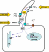
Figure 2. A working model of sensory functions of cholangiocyte cilia
See text for details. Dashed lines indicate unknown mechanisms.
Figure 3
Figure 3. Liver manifestation of ARPKD-an example of cholangiociliopathies
Light microscopy images of normal (A) and PCK (B) rat liver tissues stained with hematoxylin and eosin show healthy liver (A) and liver with large numerous cysts (B). Light microscopy image of PCK rat liver tissue stained with picrosirius red shows the presence of hepatic fibrosis around hepatic cysts (C). Scanning electron microscopy images of normal (D) and PCK (E and F) rat liver tissues show healthy liver with a conventional biliary triad (BT) consisting of the portal vein, hepatic artery and intrahepatic bile duct (D), and unhealthy liver in which cysts replace most of the liver parenchyma (E and F). A higher magnification of a portion of the liver cyst (E, the white box) is shown in F. Epithelial cells lining cysts are cholangiocytes in origin; they contain primary cilia, which are functionally abnormal due to mutation in Pkhd1encoding fibrocystin.

References

    1. Adams M, Smith UM, Logan CV, Johnson CA. Recent advances in the molecular pathology, cell biology and genetics of ciliopathies. J Med Genet. 2008 - PubMed
    1. Andrade YN, Fernandes J, Vazquez E, Fernandez-Fernandez JM, Arniges M, Sanchez TM, Villalon M, Valverde MA. TRPV4 channel is involved in the coupling of fluid viscosity changes to epithelial ciliary activity. J Cell Biol. 2005;168:869–874. - PMC - PubMed
    1. Badano JL, Mitsuma N, Beales PL, Katsanis N. The Ciliopathies: An Emerging Class of Human Genetic Disorders. Annu Rev Genomics Hum Genet. 2006;7:125–148. - PubMed
    1. Benzing T, Walz G. Cilium-generated signaling: a cellular GPS? Curr Opin Nephrol Hypertens. 2006;15:245–249. - PubMed
    1. Brailov I, Bancila M, Brisorgueil MJ, Miquel MC, Hamon M, Verge D. Localization of 5-HT(6) receptors at the plasma membrane of neuronal cilia in the rat brain. Brain Res. 2000;872:271–275. - PubMed

Publication types

MeSH terms