

Differential behavior of auricular and articular chondrocytes in hyaluronic acid hydrogels
- PMID: 18407752
- PMCID: PMC2667224
- DOI: 10.1089/ten.tea.2007.0291
Differential behavior of auricular and articular chondrocytes in hyaluronic acid hydrogels
Abstract
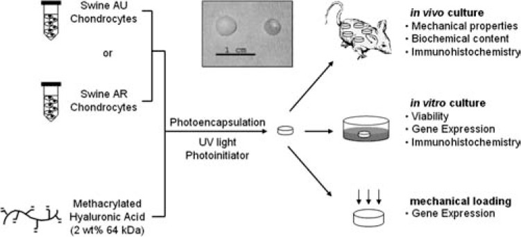
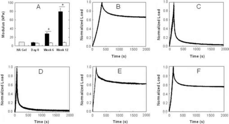
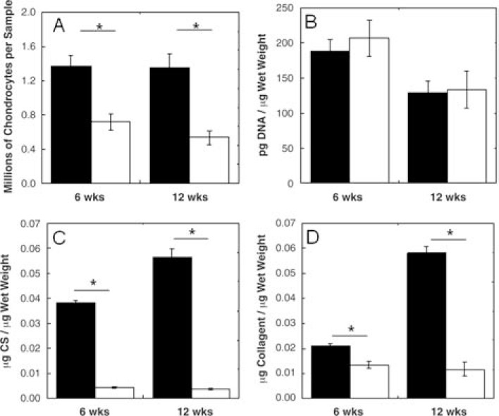
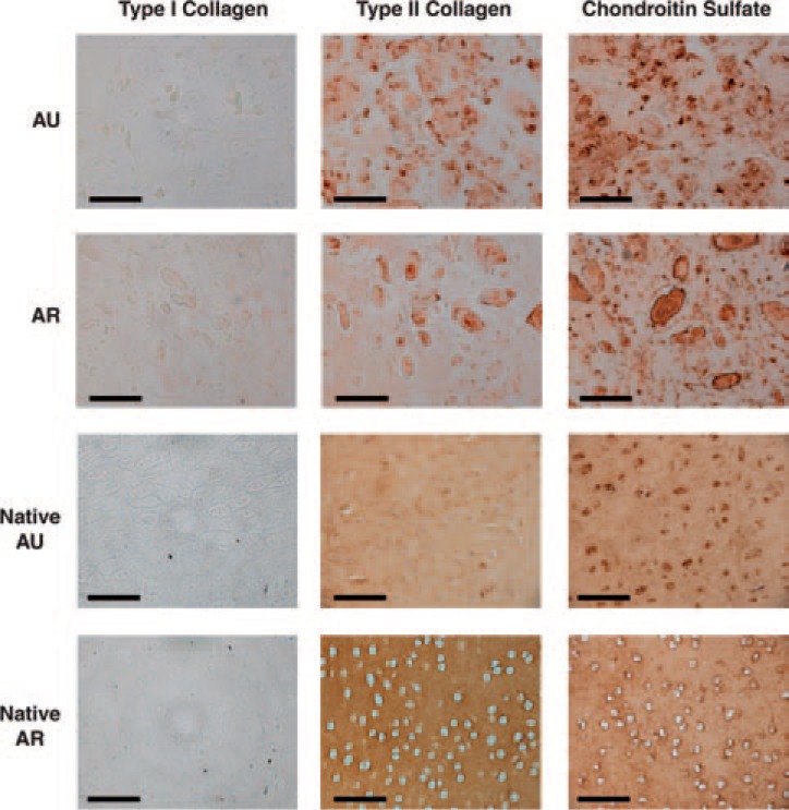
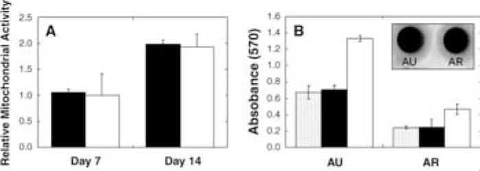
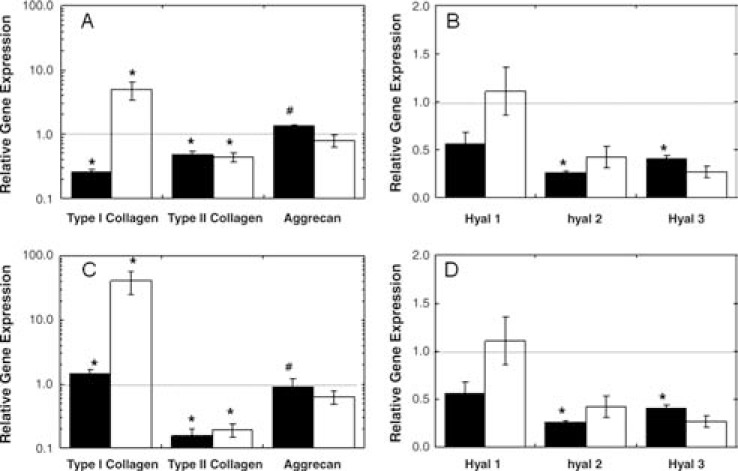
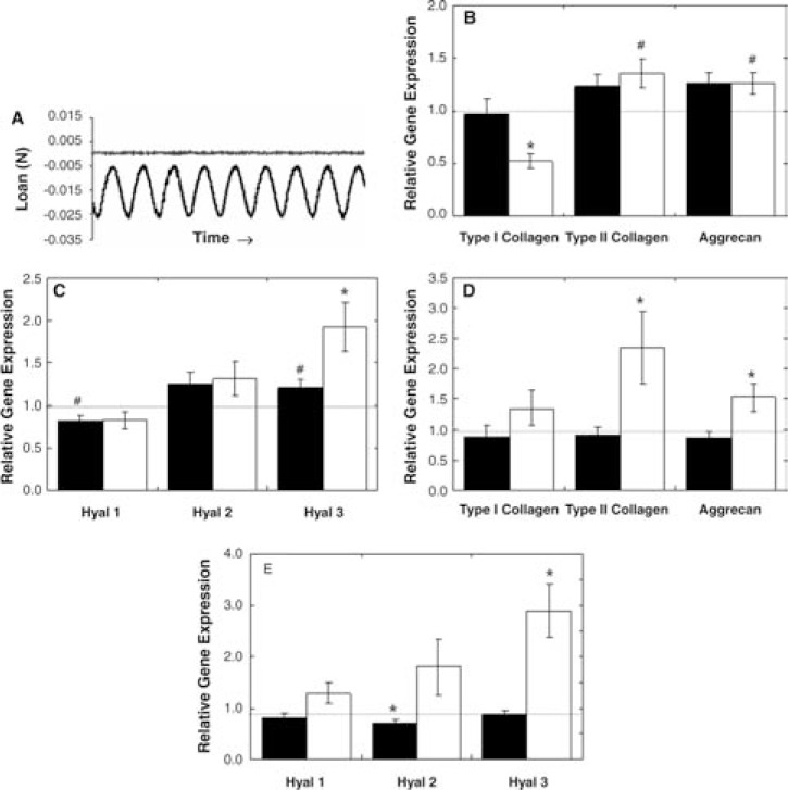
Chondrocytes isolated from a variety of sources, including auricular (AU) and articular (AR) cartilage, can differ in cell behavior, growth, and extracellular matrix (ECM) production, which can impact neocartilage properties in tissue engineering approaches. This behavior is also affected by the surrounding microenvironment, including soluble factors, biomaterials, and mechanical loading. The objective of this study was to investigate differences in juvenile AU and AR chondrocyte behavior when encapsulated in radically polymerized hyaluronic acid hydrogels. When implanted in vivo, differences in macroscopic appearance, mechanical properties, glycosaminoglycan content, and collagen content were observed depending on the chondrocyte type encapsulated. Specifically, AU constructs exhibited construct growth and neocartilage formation with increases in aggregate modulus and ECM accumulation with culture, whereas AR constructs retained their construct size and remained translucent with only a minimal increase in the compressive modulus. When cultured in vitro, both cell types remained viable and differences in gene expression were observed for type I and II collagens. Likewise, differences in gene expression were noted after dynamic mechanical loading, where stimulated AR constructs exhibited 2.3- and 1.5-fold increases in type II collagen and aggrecan over free-swelling controls, while AU samples exhibited smaller fold increases of 1.4- and 1.3-fold, respectively. Thus, these data indicate that the specific cell source, cell/material interactions, and loading environment are important in the final properties of tissue-engineered products.
Figures
References
-
- van Osch G.J.V.M., Mandl E.W., Jahr H., Koevoet W., Nolst-Trenite G., and Verhaar J.A. Considerations on the use of ear chondrocytes as donor chondrocytes for cartilage tissue engineering. Biorheology 41, 411, 2004 - PubMed
-
- Hicks D.L., Sage A.B., Schumacher B.L., Sah R.L., and Watson D. Growth and phenotype of low-density nasal septal chondrocyte monolayers. Otolaryngol Head Neck Surg 133, 417, 2005 - PubMed
-
- Kafienah W., Jakob M., Demarteau O., Frazer A., Barker M.D., Martin I., and Hollander A.P. Three-dimensional tissue engineering of hyaline cartilage: comparison of adult nasal and articular chondrocytes. Tissue Eng 8, 817, 2002 - PubMed
-
- Tay A.G., Farhadi J., Suetterlin R., Pierer G., Heberer M., and Martin I. Cell yield, proliferation, and postexpansion differentiation capacity of human ear, nasal, and rib chondrocytes. Tissue Eng 10, 762, 2004 - PubMed
-
- Panossian A., Ashiku S., Kirchhoff C.H., Randolph M.A., and Yaremchuk M.J. Effects of cell concentration and growth period on articular and ear chondrocyte transplants for tissue engineering. Plast Reconstr Surg 108, 392, 2001 - PubMed
Publication types
MeSH terms
Substances
Grants and funding
LinkOut - more resources
Full Text Sources
Other Literature Sources
Research Materials
